Application of anaplasma phagocytophilum protein APH1384
一种吞噬细胞、无形体的技术,应用在免疫学领域,能够解决检测成本高、操作时间长、价格昂贵抗原片等问题,达到提高灵敏度、快速精准诊断的效果
- Summary
- Abstract
- Description
- Claims
- Application Information
AI Technical Summary
Problems solved by technology
Method used
Image

Examples
Embodiment 1
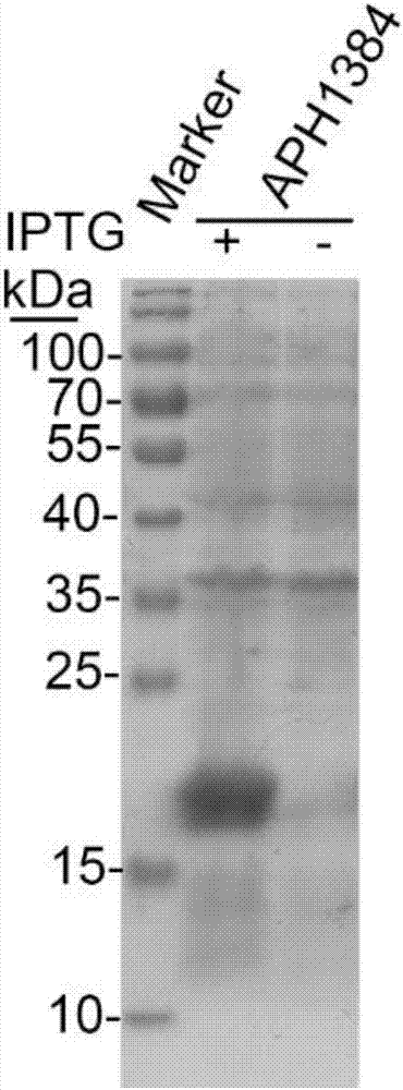
[0016] Example 1 Preparation of APH1384 protein (Anaplasma phagocytophilum species-specific protein)
[0017] Using bioinformatics technology, 610 unknown functional proteins encoded by the genome of Anaplasma phagocytophilum were targeted to screen out the species-specific proteins of Anaplasma phagocytophilum. Some of the proteins (including APH1384) were cloned, expressed and identified for their antigenicity. The results of the study found that APH1384 has strong antigenicity. The following are the detailed cloning, expression and antigenic identification steps of APH1384. Specific primers containing restriction endonuclease sites were designed according to the aph1384 gene sequence in Gene Bank: aph1384-F-TCCGAATTCATGTTTGATATATTTAATGATGCTG; aph1384-R-GCACTCGAGTTATTCGGACGCAGAGGCT. The DNA fragment of the aph1384 gene was amplified by PCR using the whole genome DNA of Anaplasma phagocytophilum as a template. The amplified DNA fragment of the target gene and the prokaryot...
Embodiment 2
[0019] Example 2 Preparation of APH1384 Polypeptide
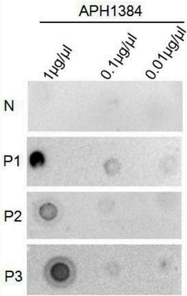
[0020] The antigenic epitope of APH1384 was predicted by bioinformatics method, and its amino acid sequence is shown in SEQ ID No.2. Chemically synthesize the APH1384 epitope polypeptide, and use DB technology to verify the antigenicity of the APH1384 polypeptide. The specific operations are as follows:
[0021] Add APH1384 polypeptide antigen dilution (1 μg / μl, 0.1 μg / μl, 0.01 μg / μl respectively) to the nitrocellulose membrane, dry at 37°C for 20-30 minutes, and block for 30 minutes. The primary antibody is the human positive serum (1: 2000) for 1 h, secondary antibody for 1 h (1:5000), and develop. see results image 3 , image 3 N in the table represents a negative serum, and P1-P3 each represent a positive serum for Anaplasma phagophilum. The results showed that the negative serum did not react with APH1384, while the three positive sera all combined with the APH1384 polypeptide, proving that the APH1384 polypeptide ...
Embodiment 3
[0022] Example 3 Application of APH1384 Polypeptide
[0023] Using the synthetic APH1384 polypeptide as the coating antigen, the ELISA method is used to detect the specific anti-APH1384 antibody in human serum. The specific operation steps are as follows:
[0024] (1) Coating: Dilute the APH1384 polypeptide synthesized in Example 2 to 20 μg / ml with PBS, and wrap it overnight at 4°C or at room temperature for 2 hours (96-well plate, 50 μl per well);
[0025] (2) Blocking: wash the 96-well plate three times with PBS, add blocking solution of bovine serum albumin (concentration: 1%), 200 μl per well, 37°C, 2h;
[0026] (3) Primary antibody incubation: Dilute the serum to be tested at a ratio of 1:1000 with blocking solution, add 100 μl of the dilution solution to each well, and incubate at 37°C for 1 hour;
[0027] (4) Secondary antibody incubation: wash four times with PBS (200 μl per well), add secondary antibody (HRP-labeled goat anti-human IgG, diluted 1:5000), and incubate ...
PUM
 Abstract
Description
Claims
Application Information
 - R&D
- Intellectual Property
- Life Sciences
- Materials
- Tech Scout
- Unparalleled Data Quality
- Higher Quality Content
- 60% Fewer Hallucinations
Browse by: Latest US Patents, China's latest patents, Technical Efficacy Thesaurus, Application Domain, Technology Topic, Popular Technical Reports.
© 2025 PatSnap. All rights reserved.Legal|Privacy policy|Modern Slavery Act Transparency Statement|Sitemap|About US| Contact US: help@patsnap.com