Pepsinogen I and pepsinogen II detection method and kit thereof

A pepsinogen and kit technology, applied in the field of biomedicine, can solve problems such as low sensitivity, inability to accurately quantify, and long detection time, and achieve the effects of improving detection sensitivity, shortening the time required for detection, and improving specificity

Active Publication Date: 2017-09-05
SHENZHEN GRADUATE SCHOOL TSINGHUA UNIV
View PDF4 Cites 7 Cited by
  • Summary
  • Abstract
  • Description
  • Claims
  • Application Information

AI Technical Summary

Problems solved by technology

Latex-enhanced immunoturbidimetric method, enzyme-linked immunoassay, and time-resolved fluorescent immunoassay, the operation process is complicated, requiring a large number of instruments and equipment and profession

Method used


Image

  • Pepsinogen I and pepsinogen II detection method and kit thereof
  • Pepsinogen I and pepsinogen II detection method and kit thereof
  • Pepsinogen I and pepsinogen II detection method and kit thereof

Examples


preparation example Construction

[0043] The general preparation method for detecting pepsinogen Ⅰ and pepsinogen Ⅱ kit includes the following steps:

[0044] (1) Preparation of fluorescent microsphere-labeled probes

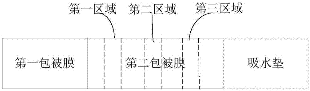
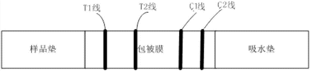
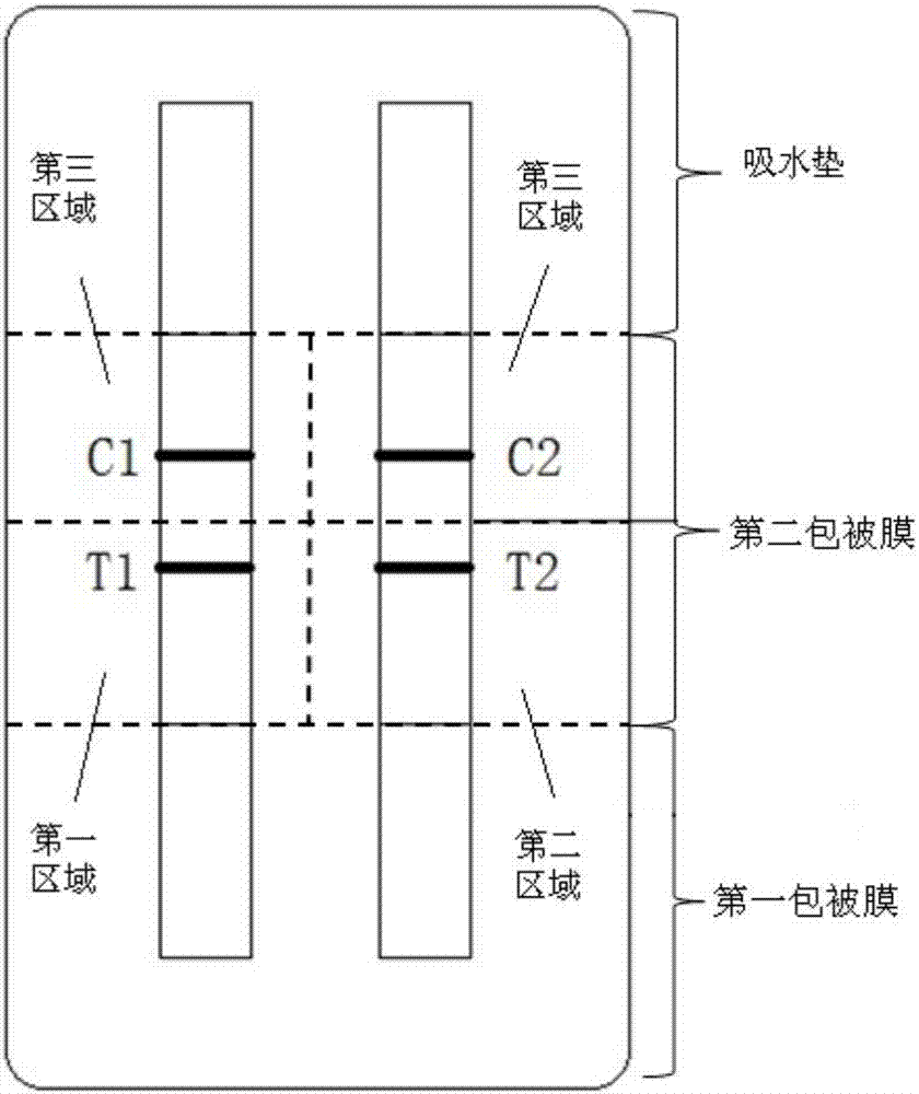
[0045] (2) Coating of antibodies at the T-line and C-line in the test area

[0046] Spray the anti-pepsinogen Ⅰ monoclonal antibody on the T1 line of the coated film test area, spray the anti-pepsinogen Ⅱ monoclonal antibody on the T2 line of the coated film test area, and spray the goat antibody on the C line by using a film spraying instrument. Mouse IgG antibody.

[0047] (3) Coating of the labeled probe at the sample pad

[0048] Spray the mixture of pepsinogen Ⅰ monoclonal antibody and pepsinogen Ⅱ monoclonal antibody labeled with fluorescent microspheres on the specific position of the sample pad by using a spraying instrument. This specific location is an area on the sample pad that serves as the subsequent "sample application end".

[0049] (4) Assembly and molding of the kit

[005...

Embodiment 1

[0057] Embodiment 1 detects the preparation of pepsinogen Ⅰ and pepsinogen Ⅱ kit

[0058] (1) Preparation of fluorescent microsphere-labeled pepsinogen Ⅰ and pepsinogen Ⅱ-labeled antibodies

[0059] Mix styrene and methyl methacrylate at a ratio of 1:1, add 1% rare earth complex Eu(TTA) 3 Phen or 0.5% CdSe / ZnS quantum dots, ultrasonically mixed to obtain liquid a. 0.05% carboxylated polyvinyl alcohol and 0.05% sodium bicarbonate were dissolved in water to obtain liquid b. Add liquid a to liquid b and ultrasonicate for 15 minutes, blow nitrogen for 30 minutes, stir to remove oxygen, and then heat to 80 degrees. Add 0.01-0.1% potassium persulfate and react for 12 hours to obtain polymer fluorescent microspheres, which are filtered, centrifuged and washed with deionized water to obtain purified functionalized fluorescent microspheres.

[0060]Take 10 mg of the above-mentioned carboxy-modified fluorescent microspheres, wash and centrifuge with MES buffer (0.1M, pH4.7), resuspen...

Embodiment 2

[0066] Example 2 Evaluation of detection kits for pepsinogen Ⅰ and pepsinogen Ⅱ

[0067] (1) Detection sensitivity

[0068] The sensitivity of the lateral flow detection reagent for detecting pepsinogen I and pepsinogen II in Example 1 was determined by using pepsinogen I and pepsinogen II antigens as samples to be tested.

[0069] At the same time, the pepsinogen Ⅰ and pepsinogen Ⅱ antigens were prepared into series concentrations (0, 0.5, 1, 5, 10, 50, 100, 500 ng / mL), were added to the sample loading end of the detection reagent for pepsinogen I and pepsinogen II obtained in Example 1, and the fluorescence intensity was detected by a fluorescence detector. Detection steps: return the sample to be tested to room temperature (25°C) before detection, and use a precise pipette to take 60 μl of the sample to be tested and drop it vertically and slowly into the lateral flow chromatography for the detection of pepsinogen Ⅰ and pepsinogen Ⅱ obtained in Example 1. Detect the samp...


PUM

PropertyMeasurementUnit
Particle sizeaaaaaaaaaa
Sensitivityaaaaaaaaaa

Abstract

The invention discloses a kit and a method for detecting pepsinogen I and pepsinogen II. The kit comprises a first coating film and a second coating film, wherein one end of the first coating film is connected with one end of the second coating film; at least one region in the first coating film is coated with a fluorescent microsphere labeled pepsinogen I antibody and a fluorescent microsphere labeled pepsinogen II antibody; the second coating film comprises a first region, a second region and a third region, which are separated from one another; a material for forming a fluorescent microsphere comprises a polystyrene-methyl methacrylate copolymer. The kit and the method, disclosed by the invention, have the advantages of high sensitivity, strong specificity, rapidness, simplicity and convenience, and can be used for realizing objectification determination and the like.

Description

technical field [0001] The invention relates to the field of biomedicine. Specifically, the present invention relates to a detection method of pepsinogen I and pepsinogen II and a kit thereof. More specifically, the present invention relates to a kit, the use of the kit in detecting pepsinogen I and pepsinogen II and a method for using the kit to detect pepsinogen I and pepsinogen II. Background technique [0002] Pepsinogen (PG), the precursor of pepsin, is divided into two subgroups based on their biochemical properties and immunogenicity. The same immunogenicity of components 1-5 is called PGⅠ, which is mainly secreted by the principal cells and mucus neck cells of the gastric glands, and components 6-7 are called PGⅡ, which are secreted by the chief cells of the oxyntic glands of the fundic mucosa of the gastric body , the mucous neck cells of the oxyntic glands, the mucous cells of the cardia glands and the pyloric glands of the gastric antrum, and the Brunner glands ...

Claims


Application Information

Patent Timeline
IPC IPC(8): G01N33/577G01N21/64
CPCG01N21/6486G01N33/577
Inventor 马岚
Owner SHENZHEN GRADUATE SCHOOL TSINGHUA UNIV
Why Eureka
Try Eureka