Accurate identification method of nucleic acid

A recognition method and nucleic acid technology, applied in the field of biometrics, can solve problems such as difficult to achieve high-resolution imaging, decrease in specific hybridization intensity, and hinder resolution, achieve stable and controllable quality of nucleic acid coatings and probes, and improve specific hybridization Intensity, reducing the effect of non-specific binding

Active Publication Date: 2021-04-16
倪燕翔
View PDF3 Cites 0 Cited by
  • Summary
  • Abstract
  • Description
  • Claims
  • Application Information

AI Technical Summary

Problems solved by technology

However, the increase in temperature leads to a decrease in the intensity of the required specific hybridization, so a higher number of imager strands has to be required to effectively label the target molecule to be detected
This prevents further improvements in resolution, making it difficult to achieve high-resolution imaging of shorter molecular regions of interest

Method used


Image

  • Accurate identification method of nucleic acid
  • Accurate identification method of nucleic acid
  • Accurate identification method of nucleic acid

Examples


Embodiment 1

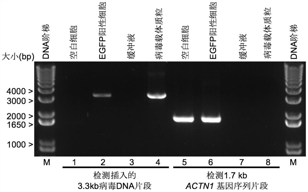
[0114] The preparation of embodiment 1 cell sample (I)

[0115] Human SK-N-SH cells were infected with the engineered pLL3.7-based lentiviral vector. After infection, cells positive for EGFP expression were screened by flow cytometry (BD FACSAria SORP Cell Sorter). In MEM medium (Gibco) containing 10% fetal bovine serum, on coverslips (Fisherbrand TM Coverglass for Growth TM Cover Glasses, Cat. No. 12-545-82) were cultured on the above-mentioned EGFP-positive cells (positive cells) or human SK-N-SH cells not infected by lentivirus (negative control).

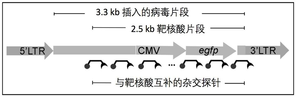
[0116] In the positive cells, a viral RNA fragment with a length of about 3.3 kb of the above-mentioned lentiviral vector was integrated into the genome of the SK-N-SH cell through reverse transcription. The integrated 3.3 kb fragment (SEQ ID NO:30) contained the sequence encoding EGFP expressed from the CMV promoter. A fragment of about 2.5 kb in length among the fragments of 3.3 kb is identified as an exemplary target nu...

Embodiment 2

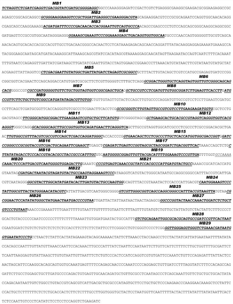
[0117] Design and synthesis of embodiment 2 hybridization probes MB1 to MB29

[0118] The exemplary hybridization probe provided in this example is a 56nt single-stranded oligonucleotide comprising a 42nt loop structure and 7nt arm structures on both sides of the loop structure. One end of the hybridization probe is conjugated with Alexa-647 as a fluorophore, and the other end is conjugated with BHQ3 as a quencher.

[0119] 29 hybridization probes were designed, denoted as MB1 to MB29, and synthesized by Life Technology. The sequences of these 29 hybridization probes are recorded in the sequence listing (SEQ ID NO: 1-29). The sequence of the loop structure is reverse complementary to a part of the antisense strand of the target nucleic acid fragment in Example 1 (that is, identical to a part of the sense strand of the target nucleic acid fragment), and 29 hybridization probes can pass through their respective loop structures The target nucleic acid fragments are labeled alon...

Embodiment 3

[0124] Embodiment 3 Hybridization probes MB1 to MB29 are specifically combined with target nucleic acid

[0125] In a buffer containing 50 mM NaCl, 1 mM EDTA, 10 mM Tris (pH 7.4), dissolve the hybridization probe, the oligonucleotide complementary to the hybridization probe (CS), and the oligonucleotide non-complementary to the hybridization probe sequence acid (NCS).

[0126] 80 nM hybridization probes (MB1 to MB29) and corresponding CS (1600 nM) or NCS (1600 nM) were reacted in 2×SSC, 50% formamide hybridization solution for 30 min at room temperature. Thereafter, fluorescence emission readings were taken at 665 nm using a spectrofluorometer excited with a 647 nm laser. Readouts for each hybridization probe are shown in Figure 3a . For each hybridization probe, the fluorescence emission readout after co-reaction with CS was significantly higher than that after co-reaction with NCS, indicating the specific binding of each hybridization probe to the target sequence.

[01...


PUM


Abstract

The present disclosure relates to a nucleic acid identification method. The identification method comprises the steps of: a. contacting the sample with a hybridization probe, b. optionally, eluting the hybridization probe not bound to the target nucleic acid, and c. identifying whether the hybridization probe is bound to the target nucleic acid binding; wherein the hybridization probe comprises a reporter group, and the state of the hybridization probe is different in the following two cases, so that a recognizable signal is generated by the reporter group: (i) the hybridization probe The needle is free and (ii) the hybridization probe is bound to the target nucleic acid. The present disclosure also relates to the use of hybridization probes for fluorescent microscopic imaging of target nucleic acids. The disclosed identification method can simplify the operation steps of nucleic acid identification, efficiently label target nucleic acid molecules, reduce the non-specific binding between probes and nucleic acids other than target nucleic acids, improve the efficiency and accuracy of identification, and is especially beneficial for short length and and / or identification of non-repetitive nucleic acids, suitable for obtaining super-resolution images of target nucleic acids.

Description

technical field [0001] The present disclosure relates to the field of biometric identification, in particular to a method for precise nucleic acid identification. Background technique [0002] In order to study the structure and interaction of nucleic acid molecules in cells, it is necessary to identify the presence, position, and shape of nucleic acid molecules. A common method is fluorescence in situ hybridization (fluorescence in situ hybridization, FISH). In this method, the probe and the target nucleic acid are hybridized based on the high degree of complementarity of the sequence, and then imaged by means of a fluorophore that directly or indirectly labels the probe, thereby identifying and locating the target nucleic acid of a specific sequence (Non-Patent Document 1 ). [0003] However, due to the limitation of the diffraction limit, traditional optical microscopy can only obtain imaging resolutions of about 200-300 nm in the lateral direction and about 600 nm in t...

Claims


Application Information

Patent Timeline
Patent Type & Authority Patents(China)
IPC IPC(8): C12Q1/6816G01N21/64
CPCC12Q1/6841G01N21/6486C12Q2563/107
Inventor 倪燕翔牛钢马紫珊曹博张奇伟
Owner 倪燕翔
Why Eureka
Try Eureka