Intelligent and automatic delineation method for gross tumor volume and organs at risk

A tumor radiation and treatment target technology, applied in the field of biomedical engineering, can solve the problems of strong subjectivity, affecting the accuracy of radiation treatment planning, treatment efficacy, and poor precision

Inactive Publication Date: 2017-11-28
强深智能医疗科技(昆山)有限公司
View PDF2 Cites 171 Cited by
  • Summary
  • Abstract
  • Description
  • Claims
  • Application Information

AI Technical Summary

Problems solved by technology

[0004] The purpose of the present invention is to provide an intelligent and automatic delineation method for tumor radiotherapy targets and organs at risk, so as to solve the problem of low efficiency, strong subjectivity, and poor accuracy of manual target delineation in clinical practice, which ultimately affects the accuracy of radiotherapy planning and treatment. Efficacy and other issues

Method used


Image

  • Intelligent and automatic delineation method for gross tumor volume and organs at risk
  • Intelligent and automatic delineation method for gross tumor volume and organs at risk
  • Intelligent and automatic delineation method for gross tumor volume and organs at risk

Examples


Embodiment 1

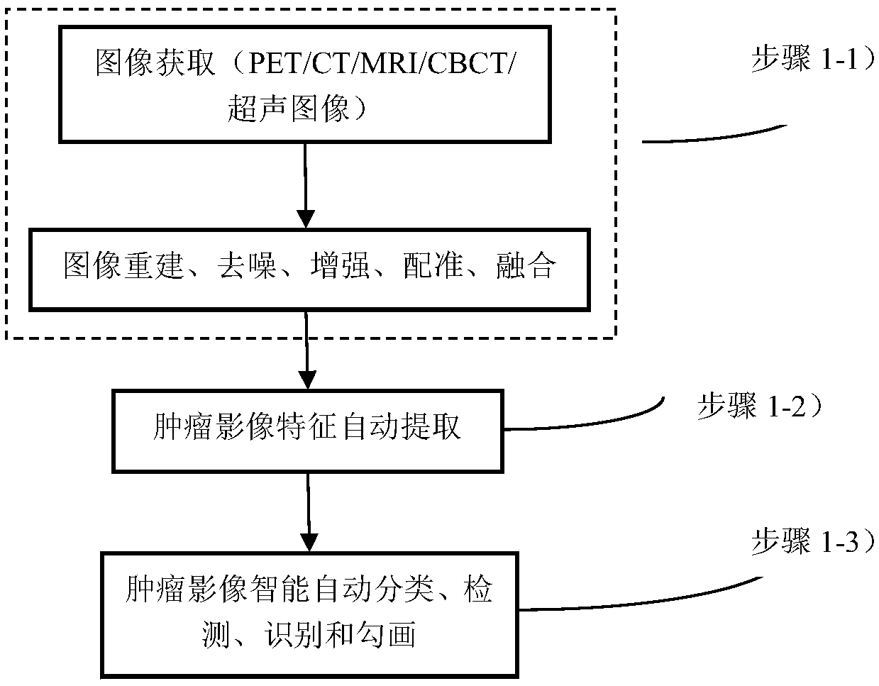
[0068] This embodiment shows an image preprocessing method, including image acquisition and preprocessing of the acquired graphics:

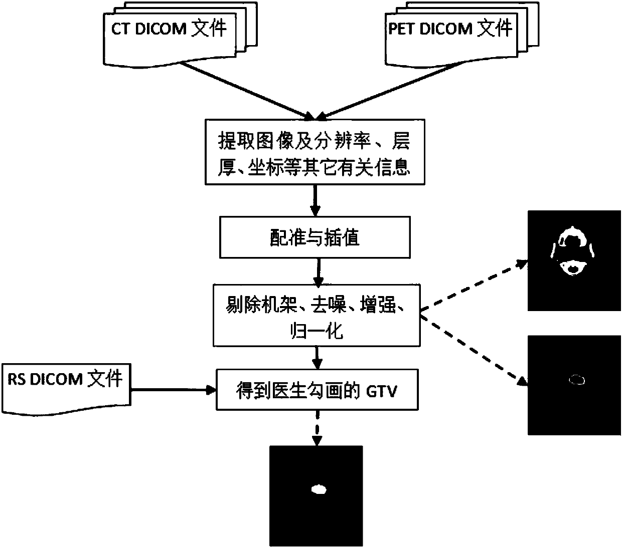
[0069] Image acquisition includes acquiring images of two parts: (a) PET / CT, PET / MRI, MRI, CT and other images for tumor diagnosis, and radiotherapy simulation positioning CT (SCT) for making treatment plans. These images are commercially imaged by the hospital Equipment imaging is obtained through the DICOM file exported by the PACS system in the hospital clinic, and the DICOM file also provides scanning parameter information. (b) Onboard cone-beam CT (CBCT), MRI, or ultrasound-guided images for radiotherapy guidance.

[0070] The image information of various modalities is preprocessed, and SCT is used as a reference image to register and interpolate the images of each modality into images with the same spatial resolution as SCT. Specifically, tumor PET / CT / MRI medical imaging systems have different imaging principles and characteristics, and p...

Embodiment 2

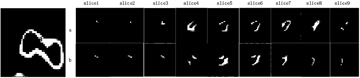
[0076] This example shows a random walk tumor delineation method integrating an adaptive regression kernel:

[0077] The traditional random walk method only considers the gray information of the image, and uses the edge weight function of the undirected graph to represent the similarity between adjacent pixels in space, without considering the neighborhood information of the pixel; while the PET image of the tumor It has obvious anisotropy characteristics, mainly manifested in the uneven distribution of SUV values ​​in the tumor area; in PET images, the SUV (Standard Uptake Value) value is affected by the intensity of tissue metabolism, and the more active the metabolism, the higher the corresponding SUV value; The metabolism of normal brain tissue around head and neck tumors is also active, and its SUV value distribution is close to that of the tumor area, and the edge weight function is close to 1. At this time, the random walk algorithm cannot effectively distinguish them. ...

Embodiment 3

[0114] This example shows a method for delineating organs at risk and tumors based on a deep neural network learning method: in the process of clinical radiotherapy planning for malignant tumors, not only must the malignant tumors be accurately delineated to achieve highly conformal tumor irradiation, but also it is necessary to The normal tissues and organs adjacent to the tumor are delineated with high precision to avoid irradiation of normal tissues and organs to the greatest extent. Using deep learning methods, it automatically learns from tumor PET / CT / MRI images to obtain deep-level, high-level, and implicit specific features of tumor radiotherapy target areas and organs at risk. Based on these features, high-precision delineation of organs at risk and malignant tumor targets can improve the accuracy of delineation of organs at risk and tumors. In this example, all clinically available tumor CT, PET, and MRI images are used to detect, classify, identify, and delineate tum...


PUM


Abstract

The invention relates to an intelligent and automatic delineation method for a gross tumor volume and organs at risk, which includes the following steps: (1) tumor multi-modal (mode) image reconstruction, de-noising, enhancement, registration, fusion and other preprocessing; (2) automatic extraction of tumor image features: automatically extracting one or more pieces of tumor image group (texture feature spectrum) information from CT, CBCT, MRI, PET and/or ultrasonic multi-modal (mode) tumor medical image data; and (3) intelligently and automatically delineating a gross tumor volume and organs at risk through deep learning, machine learning, artificial intelligence, regional growing, the graph theory (random walk), a geometric level set, and/or a statistical theory method. A gross tumor volume (GTV) and organs at risk (OAR) can be delineated with high precision.

Description

technical field [0001] The present invention relates to technical fields such as medical imaging, medical image analysis and processing, deep learning, machine learning, big data analysis, artificial intelligence and tumor radiophysics, radiobiology, radiotherapy, biomedical engineering, etc.; The method of image features and the method of intelligent and automatic classification, detection, identification, and segmentation of tumor radiotherapy target volume and radiotherapy endangered organs; specifically, it demonstrates the intelligent and automatic delineation method of tumor radiotherapy target volume and endangered organs. Background technique [0002] Tumor radiation therapy is one of the three major tumor treatment techniques at present. Accurate radiotherapy for malignant tumors relies on computed tomography (CT), magnetic resonance imaging (MRI), positron emission tomography (PET), cone beam computed tomography (CBCT) technologies and corresponding intelligent pro...

Claims


Application Information

Patent Timeline
Patent Type & Authority Applications(China)
IPC IPC(8): G06K9/62G06T15/00G06T7/33G06T5/00G06N3/04
CPCG06T5/002G06T7/33G06T15/00G06T2207/20221G06T2207/30096G06T2207/20081G06T2207/10104G06T2207/10088G06T2207/10081G06V2201/032G06N3/045G06F18/241G06F18/253
Inventor 刘国才田娟秀官文静
Owner 强深智能医疗科技(昆山)有限公司
Why Eureka
Try Eureka