Fluorescent bleaching-based super-resolution imaging method

A super-resolution imaging and fluorescence bleaching technology, which is applied in the field of super-resolution imaging based on fluorescence bleaching, can solve the problems of high technical requirements, low universality, and insufficient resolution of ordinary optical microscopes, and achieve resolution improvement and cost reduction , the effect of resolution improvement

Inactive Publication Date: 2017-12-01
SHANGHAI INST OF APPLIED PHYSICS - CHINESE ACAD OF SCI
View PDF2 Cites 5 Cited by
  • Summary
  • Abstract
  • Description
  • Claims
  • Application Information

AI Technical Summary

Problems solved by technology

[0005] The purpose of the present invention is to provide a super-resolution imaging method based on fluorescence bleaching, so as to solve the problem that the resolution of ordinary optical microscopes in the prior art is not enough, and most existing super-resolution fluorescence microscopes have too high requirements for equipment and technology. less common problems

Method used


Image

  • Fluorescent bleaching-based super-resolution imaging method
  • Fluorescent bleaching-based super-resolution imaging method
  • Fluorescent bleaching-based super-resolution imaging method

Examples


Embodiment 1

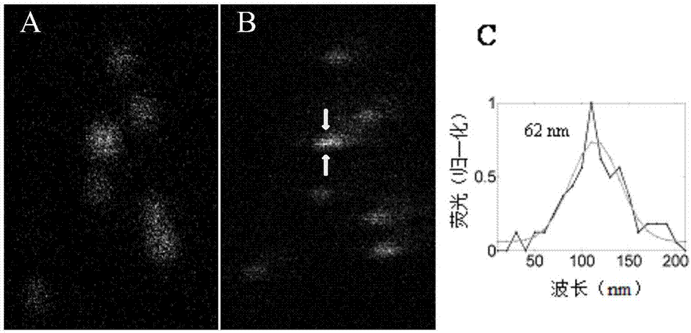
[0040] Super-resolution imaging of Alexa405-labeled fluorescent beads.

[0041] 1.1 Alexa405 DNA single strand (sequence: Alexa 405-5'AAAAAACAGGACCAGAAAAAA-3'biotin, TAKARA) was diluted to 100 μM with ultrapure water.

[0042] 1.2 Dilute the polystyrene beads (Invitrogen) labeled with bioavidin to 100 μM with ultrapure water.

[0043] 1.3 Add 97 μL of PBS to the 100 μL centrifuge tube, and add 2 μL of the DNA single strand just diluted and 1 μL of polystyrene beads labeled with bioavidin. This was incubated at 25°C for 1 hour.

[0044] 1.4 Add the incubated solution into a 100KD ultrafiltration tube (Millipore), and then add 300 microliters of PBS.

[0045] 1.5 Put it into a centrifuge and centrifuge at a speed of 2700g for 2 minutes. After taking out the centrifuge tube, pour off the filtered solution, then add 400 microliters of PBS to the centrifuge tube, and centrifuge at a speed of 2700g. After repeating three times, remove the centrifuged liquid.

[0046] 1.6 Put th...

Embodiment 2

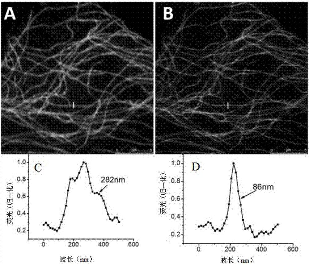
[0052] Example 2: Super-resolution imaging of microtubules labeled with Alexa 405 by fluorescence bleaching

[0053] 2.1 Put in the 12-well cell culture plate 18mm round coverslip, on which approximately 60,000 Hela cells were plated.

[0054] 2.2 Wash 3 times with 1 mL of 1× phosphate buffered saline (PBS) at 37°C, with an interval of five minutes between each time.

[0055] 2.3 Add 1 mL of 4% paraformaldehyde (w / v) + 4% sucrose (w / v) to fix for 15 min.

[0056] 2.4 Wash 3 times with 1×PBS buffer, with an interval of 5 minutes each time.

[0057] 2.5 Dilute Triton X-100 to 0.25% (w / v) with 6% bovine serum albumin (w / v). Add 500 μL 0.25% Triton X-100 to the twelve-well plate and incubate for 45 minutes.

[0058] 2.6 Add 400mL primary antibody (10nM) and incubate for 1h.

[0059] 2.7 Wash three times with 1×PBS buffer, with an interval of 10 minutes each time.

[0060] 2.8 Add 400mL of secondary antibody labeled with Alexa 405 and incubate for 45min.

[0061] 2.9 Wash w...

Embodiment 3

[0064] Example 3: Ultra-resolution imaging of Atto 488-labeled cellular microtubules by fluorescence bleaching

[0065] 3.1 Put a φ18mm round cover slip in a 12-well cell culture plate, and spread about 60,000 Hela cells on the slip.

[0066] 3.2 Wash 3 times with 1 mL of 1× phosphate buffered saline (PBS) at 37°C, with an interval of five minutes between each time.

[0067] 3.3 Add 1 mL of 4% paraformaldehyde (w / v) + 4% sucrose (w / v) to fix for 15 minutes.

[0068] 3.4 Wash 3 times with 1×PBS buffer, with an interval of 5 minutes each time.

[0069] 3.5 Dilute Triton X-100 to 0.25% (w / v) with 6% bovine serum albumin (w / v). Add 500 μL 0.25% Triton X-100 to the twelve-well plate and incubate for 45 minutes.

[0070] 3.6 Add 400mL primary antibody (10nM) and incubate for 1h.

[0071] 3.7 Wash with 1×PBS buffer three times, with an interval of 10 minutes each time.

[0072] 3.8 Add 400mL of secondary antibody labeled Atto 488 and incubate for 45min.

[0073] 3.9 Wash with 1...


PUM


Abstract

The invention provides a fluorescent bleaching-based super-resolution imaging method. The fluorescent bleaching-based super-resolution imaging method comprises the following steps of 1) marking a sample through fluorescent dye with photo-bleaching characteristics; 2) focusing laser beams onto the marked sample to be tested through a point excitation scanning type laser microscope, wherein the sample to be tested emits fluorescence under laser beam excitation; 3) collecting the fluorescence emitted by the sample to be tested to acquire laser intensity information; 4) scanning the sample point by point through exciting light of certain power at certain scanning parameters so as to directly bleach the sample during the scanning process, to relatively reduce the number of collected effective pixels, to further obtain relatively small fluorescence spots and to directly achieve super-resolution images breaking through optical diffraction limits.

Description

technical field [0001] The invention relates to the field of super-resolution far-field optical imaging that breaks through the optical diffraction limit, and more specifically relates to a super-resolution imaging method based on fluorescence bleaching. Background technique [0002] Optical microscopes can non-destructively observe and image biological samples in real time, and are essential tools in biological research. The development of optical microscopes also promotes the progress of life sciences. The organelles and proteins in the cell are all at the nanoscale, which requires the resolution of the microscope to reach the nanoscale to be able to distinguish them clearly. However, due to the existence of optical diffraction, the lateral resolution of the optical microscope is limited to about 200nm, which hinders people from using the optical microscope to conduct more in-depth research. [0003] In the last two decades, a series of super-resolution microscopes have ...

Claims


Application Information

Patent Timeline
Patent Type & Authority Applications(China)
IPC IPC(8): G01N21/64
CPCG01N21/6428G01N21/6456G01N2021/6432G01N2021/6439
Inventor 樊春海邓素辉程亚陈建芳侯尚国
Owner SHANGHAI INST OF APPLIED PHYSICS - CHINESE ACAD OF SCI
Why Eureka
Try Eureka