Method for separating components in mixture by use of elisa plate and monoclonal antibody

A technology of monoclonal antibody and separation method, which is applied in the field of separation of components in the mixture by using microplate and monoclonal antibody, which can solve the problems of large influence of experimental conditions, quantitative detection, and insufficient specificity, and shorten the detection time , Reduce detection cost, improve detection accuracy

Active Publication Date: 2017-12-05
云南沃森生物技术股份有限公司
View PDF5 Cites 2 Cited by
  • Summary
  • Abstract
  • Description
  • Claims
  • Application Information

AI Technical Summary

Problems solved by technology

There are in this method: 1. Insufficient specificity: the deficiency of poor effect on the separation of components with similar molecular size and properties
2. It is greatly affected by the experimental conditions: the concentration of the solution, the pH value, etc.
4. The required experimental steps, equipment and experimental time are relatively long
[0026] At present, the existing technology can only detect the content of total polysaccharide or total protein carrier in the conjugate vaccine by ELISA method, and cannot directly quantitatively detect the polysaccharide-protein carrier conjugate, free polysaccharide and free protein in the conjugate vaccine respectively

Method used


Image

  • Method for separating components in mixture by use of elisa plate and monoclonal antibody
  • Method for separating components in mixture by use of elisa plate and monoclonal antibody
  • Method for separating components in mixture by use of elisa plate and monoclonal antibody

Examples


Embodiment 1

[0060] Example 1: Detection of free A content in A-TT conjugate vaccine

[0061] 1.1 Source of antigen

[0062] The stock solution of A-TT conjugate was prepared according to "Group A and Group C Meningococcal Polysaccharide Conjugate Vaccine" in "Chinese Pharmacopoeia 2015 Edition".

[0063] 1.2 Sources of monoclonal antibodies

[0064] Group A meningococcal polysaccharide monoclonal antibody: prepared by the hybridoma cell line WV-A-01 (CCTCC NO: C2015229, China Center for Type Culture Collection) according to the mouse monoclonal antibody ascites preparation method.

[0065] Tetanus toxoid (TT) monoclonal antibody: prepared from hybridoma cell line WV-TT-05 (CCTCC NO: C2014200, China Center for Type Culture Collection) according to the mouse monoclonal antibody ascites preparation method.

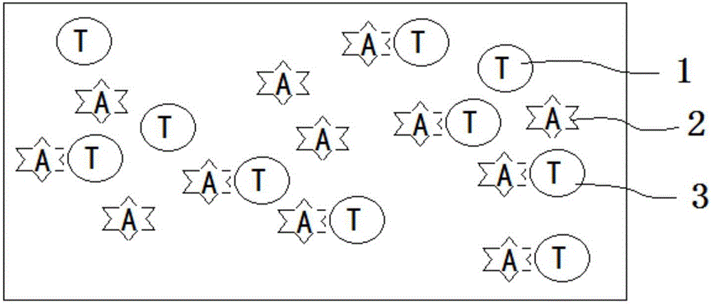
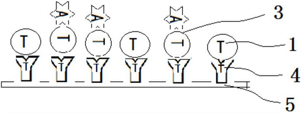
[0066] 1.3 Separation of free group A polysaccharides in A-TT conjugate vaccines using TT monoclonal antibody

[0067] 1.3.1 Coat 50 ng / well of monoclonal antibody against tetanus tox...

Embodiment 2

[0084] Embodiment 2: the detection of free TT content in A-TT conjugate vaccine

[0085] 1.1 Source of antigen

[0086] The stock solution of A-TT conjugate was prepared according to "Group A and Group C Meningococcal Polysaccharide Conjugate Vaccine" in "Chinese Pharmacopoeia 2015 Edition".

[0087] 1.2 Sources of monoclonal antibodies

[0088] Group A meningococcal polysaccharide monoclonal antibody: prepared by the hybridoma cell line WV-A-01 (CCTCC NO: C2015229, China Center for Type Culture Collection) according to the mouse monoclonal antibody ascites preparation method.

[0089] Tetanus toxoid (TT) monoclonal antibody: prepared from hybridoma cell line WV-TT-05 (CCTCC NO: C2014200, China Center for Type Culture Collection) according to the mouse monoclonal antibody ascites preparation method.

[0090] 1.3 Isolation of free TT in A-TT conjugate vaccine using A monoclonal antibody

[0091] 1.3.1 Coat 50ng / well of group A meningococcal monoclonal antibody on an ELISA pl...


PUM


Abstract

The present invention discloses a method for separating components in a mixture by use of an elisa plate and a monoclonal antibody. The separation method comprises the following steps: the elisa plate is coated with an antibody of a component A in the mixture, substances containing the component A in the mixture to be tested are bonded to form an antigen-antibody complex immobilized on the plate, supernatant contains no A component, so that components excluding the component A can be separated out. The method is more precise and specific for separating the components. Only one same detection system can separate and quantitatively determine different substances to be determined containing one same component. The volume of the needed sample to be tested is greatly reduced from milliliters to microliters; complicated equipment investments are not required, testing cost is reduced, testing time is greatly shortened from 3 to 4 days to 3 to 4 hours, and detection accuracy is greatly increased from micrograms to nanograms.

Description

technical field [0001] The invention belongs to the technical field of material separation, and in particular relates to a method for separating components in a mixture by using an enzyme label plate and a monoclonal antibody. Background technique [0002] In 1971, Engvall and Perlmann published an article on enzyme-linked immunosorbent assay (enzyme linkedimmunosorbent assay, ELISA) for the quantitative determination of IgG, which made the enzyme-labeled antibody technology used for antigen localization in 1966 develop into the determination of trace substances in liquid specimens method. The basic principle of this method is: ① Make the antigen or antibody bind to the surface of a certain solid phase carrier and maintain its immunological activity. ②The antigen or antibody is connected with an enzyme to form an enzyme-labeled antigen or antibody, which not only retains its immune activity, but also retains the activity of the enzyme. During the determination, the test sp...

Claims


Application Information

Patent Timeline
Patent Type & Authority Applications(China)
IPC IPC(8): G01N33/535G01N33/577G01N33/543
CPCG01N33/535G01N33/54306G01N33/577
Inventor 钱雯陈玉秋王丽丽施競陈南萍熊应景
Owner 云南沃森生物技术股份有限公司
Why Eureka
Try Eureka