Method for isolating, culturing and differentiating neural crest stem cells from DRG (dorsal root ganglion)

A technology of dorsal root ganglion and neural crest, applied in the field of stem cell biology, can solve the problems of large cell loss and incomplete enteric nervous system, and achieve the effect of strong proliferation ability

Active Publication Date: 2017-12-15
ZHEJIANG UNIV
View PDF3 Cites 4 Cited by
  • Summary
  • Abstract
  • Description
  • Claims
  • Application Information

AI Technical Summary

Problems solved by technology

In recent years, there have been many studies on the isolation and culture of neural crest stem cells derived from dorsal root ganglion, mainly using the method of flow cytometry to separate cells. This method can obtain relatively pure cell sources, but the loss of cells is relatively large
[0004] At present, there ar

Method used


Image

  • Method for isolating, culturing and differentiating neural crest stem cells from DRG (dorsal root ganglion)
  • Method for isolating, culturing and differentiating neural crest stem cells from DRG (dorsal root ganglion)
  • Method for isolating, culturing and differentiating neural crest stem cells from DRG (dorsal root ganglion)

Examples


Embodiment 1

[0030] Example 1, Isolation and purification of neural crest stem cells derived from dorsal root ganglion

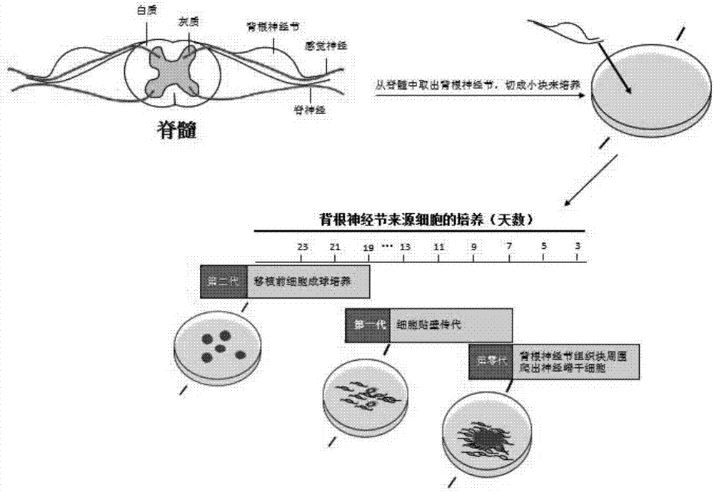
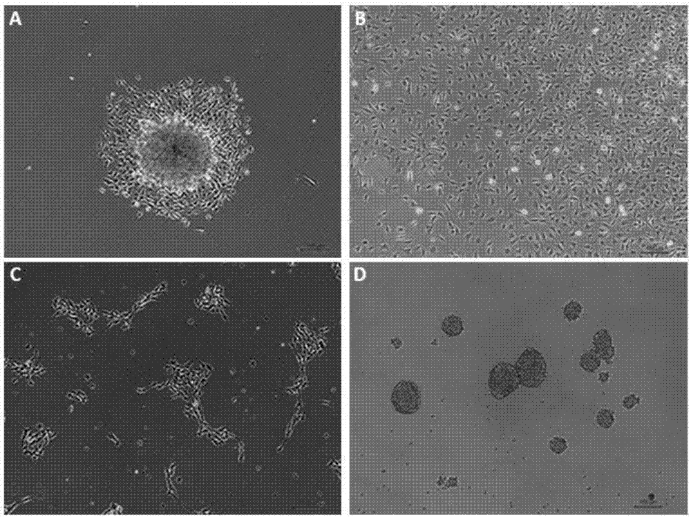
[0031] Take out the C57 mice about 6 days after birth, soak them in alcohol for 15 minutes, incise the back under the ultra-clean table, take out the neural tube, and take out the dorsal root ganglion at the edge of the neural tube under a microscope, cut it into pieces of about 2 mm, After rinsing with PBS, centrifuge at 800rmp for 5 minutes to collect the tissue pieces, resuspend the tissue pieces in the primary medium, and transfer them to poly-ornithine-coated plates, such as figure 1 shown in the flow chart. The primary medium is: DMEM / F12 medium, 1% N2, 2% B27, 20ng / ml bFGF (basic fibroblast growth factor), 20ng / ml EGF (endothelial growth factor), 100IU / L penicillin / streptomycin. Placed in a 37-degree constant temperature incubator containing 5% CO2 for cultivation. After two days in culture, cells crawled out around the tissue block, as figure 2 ‐As shown i...

Embodiment 2

[0032] Example 2, identification of neural crest stem cells derived from dorsal root ganglia.

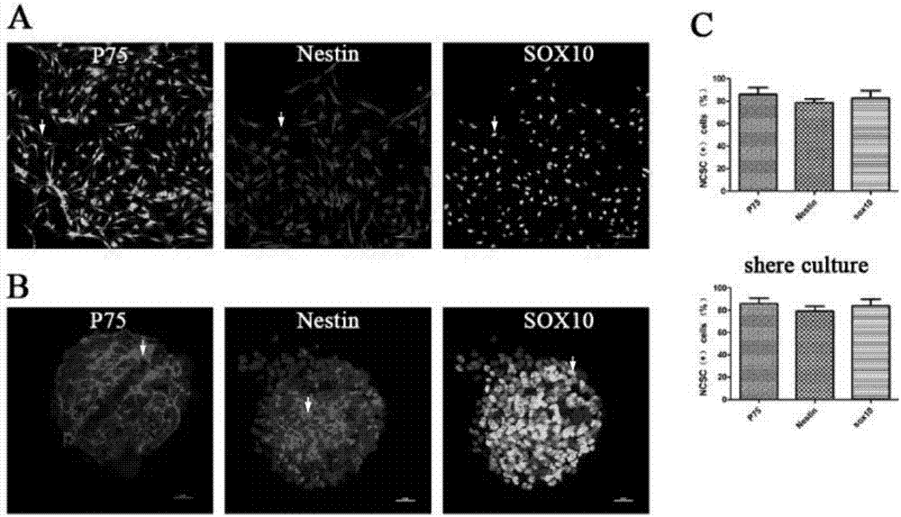
[0033] Take the cells that have been cultured for 2 passages and sphere culture, rinse twice with PBS, fix with paraformaldehyde for 15 minutes, block with blocking solution (1% BSA) containing 0.3% (volume ratio) triton for 30 minutes at room temperature, and work with the primary antibody diluted 1:100 Solution (P75, Nestin, SOX10) was incubated at room temperature for 2 hours, washed three times with PBS, incubated with secondary antibody dilution (1:500) for one hour at room temperature, rinsed three times with PBS, observed and photographed under a fluorescence microscope, and counted by image J software The proportion of positive cells. Such as image 3 As shown, the neural crest stem cells derived from the dorsal root ganglion obtained by the method of the present invention have relatively high purity, and more than 90% of the neural crest stem cells derived from the dorsal r...

Embodiment 3

[0034] Example 3, analysis of neural crest stem cell expansion ability derived from dorsal root ganglion

[0035] The neural stem cells obtained from the primary generation are subcultured and expanded in two forms, one is single-layer adherent culture, and the other is spheroid suspension culture, such as figure 2 (C-D) Both cultures shown in (C-D) can be expanded for several generations in vitro.

[0036] The cells of the first and fifth passages of adherent culture were taken, rinsed twice with PBS, fixed with 4% (mass ratio) paraformaldehyde for 15 minutes, blocked with 0.3% triton blocking solution (1% BSA) at room temperature for 30 minutes, 1 :200 diluted Ki67 primary antibody working solution was incubated at room temperature for 2 hours, washed three times with PBS, incubated with secondary antibody dilution (1:500) for 1 hour at room temperature, rinsed three times with PBS, observed and photographed under a fluorescence microscope, through image J The software cou...


PUM


Abstract

The invention provides a novel method for isolating, culturing and differentiating neural crest stem cells from DRG (dorsal root ganglion). The method comprises steps as follows: (1) primary isolation of the neural crest stem cells from DRG; (2) purification and culture amplification of the neural crest stem cells from DRG; (3) induced differentiation of the neural crest stem cells from DRG. The obtained neural crest stem cells have higher multiplication capacity and still have multiplication capacity after being continuously cultured for generations in vitro.

Description

technical field [0001] The invention belongs to the technical field of stem cells, and relates to methods for separating, cultivating and differentiating neural crest stem cells derived from dorsal root ganglia. Background technique [0002] Neural crest stem cells are an important cell type during embryonic development. Originating from the dorsal portion of the neural tube during early embryonic development, it differentiates into various cell types by migrating to various parts of the embryo. The neural crest cells are mainly divided into cranial-derived neural crest cells and trunk neural crest stem cells. The trunk-derived neural crest cells are divided into vagus neural crest and sacral neural crest. After the neural crest cells migrate to various parts of the body, they can differentiate into melanocytes, Craniofacial cells, chondrocytes, bone cells, smooth muscle cells, central and peripheral neurons, and glial cells. [0003] The dorsal root ganglion (DRG) is form...

Claims


Application Information

Patent Timeline
IPC IPC(8): C12N5/0797C12N5/0793
CPCC12N5/0619C12N5/0623C12N2500/24C12N2500/32C12N2500/38C12N2500/40C12N2500/44C12N2501/105C12N2501/11C12N2501/115C12N2501/13C12N2501/999C12N2533/32C12N2533/52
Inventor 陈伟胡珲丁园园李颖
Owner ZHEJIANG UNIV
Why Eureka
Try Eureka