Pathological tissue specimen fixative
A tissue specimen and fixative technology, applied in the field of specimen processing, can solve the problems of specimen dehydration, high cost, deformation, etc., and achieve the effect of stable performance, safe use, and less dosage
- Summary
- Abstract
- Description
- Claims
- Application Information
AI Technical Summary
Problems solved by technology
Method used
Image
Examples
Embodiment 1
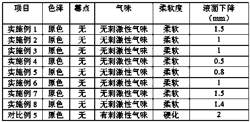
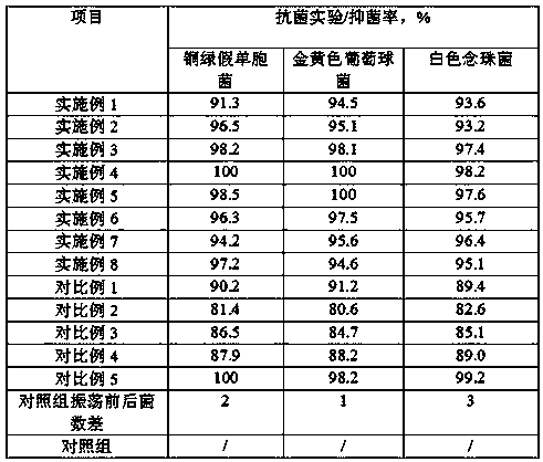
[0020] A fixative solution for pathological tissue specimens, comprising the following components: 10 mL of glycerol, 32 g of sodium chloride, 0.5 g of berberine, 1.2 g of menthol, 8 g of sodium dichloroisocyanurate, 2 mL of dimethyl sulfoxide, and a film-forming assistant Agent 6g, alkylphenol polyoxyethylene ether 0.5g, pure water to 1000mL.
[0021] The film-forming aid is formed by mixing sodium alginate and chitosan at a weight ratio of 1:1.
[0022] The pathological tissue specimen fixative is prepared by the following steps:
[0023] Weigh sodium chloride and 500mL pure water and mix evenly to obtain sodium chloride solution, then add berberine and dimethyl sulfoxide mixture in sequence, stir at a high speed of 1500r / min for 15min, then add glycerol, menthol, Sodium dichloroisocyanurate, film-forming aid, alkylphenol polyoxyethylene ether, and pure water were adjusted to 1000mL, and stirred at a low speed of 500r / min for 35min to obtain the product.
Embodiment 2
[0025] A pathological tissue specimen fixative, comprising the following components: glycerol 11mL, sodium chloride 31g, berberine 0.6g, menthol 1.1g, sodium dichloroisocyanurate 9g, dimethyl sulfoxide 2.2mL, film-forming 5g of additives, 0.6g of alkylphenol polyoxyethylene ether, and dilute to 1000mL with pure water.
[0026] The film-forming aid is formed by mixing sodium alginate and chitosan at a weight ratio of 1:1.
[0027] The pathological tissue specimen fixative is prepared by the following steps:
[0028] Weigh sodium chloride and 500mL pure water and mix evenly to obtain sodium chloride solution, then add berberine and dimethyl sulfoxide mixture successively, stir at a high speed of 1600r / min for 10min, then add glycerin, menthol, Sodium dichloroisocyanurate, film-forming aids and alkylphenol polyoxyethylene ethers were adjusted to a volume of 1000mL, and stirred at a low speed of 600r / min for 25min to obtain the product.
Embodiment 3
[0030] A fixative solution for pathological tissue specimens, comprising the following components: 12 mL of glycerol, 30 g of sodium chloride, 0.7 g of berberine, 1.0 g of menthol, 10 g of sodium dichloroisocyanurate, 2.4 mL of dimethyl sulfoxide, and Auxiliary 4g, alkylphenol polyoxyethylene ether 0.7g, pure water to 1000mL.
[0031] The film-forming aid is formed by mixing sodium alginate and chitosan at a weight ratio of 1.5:1.
[0032] The pathological tissue specimen fixative is prepared by the following steps:
[0033] Weigh sodium chloride and 500mL pure water and mix evenly to obtain sodium chloride solution, then add berberine and dimethyl sulfoxide mixture in sequence, stir at a high speed of 1700r / min for 14min, then add glycerol, menthol, Sodium dichloroisocyanurate, film-forming aids, alkylphenol polyoxyethylene ether, and pure water were adjusted to 1000mL, and stirred at a low speed of 700r / min for 30min to obtain the product.
PUM
 Abstract
Description
Claims
Application Information
 - R&D
- Intellectual Property
- Life Sciences
- Materials
- Tech Scout
- Unparalleled Data Quality
- Higher Quality Content
- 60% Fewer Hallucinations
Browse by: Latest US Patents, China's latest patents, Technical Efficacy Thesaurus, Application Domain, Technology Topic, Popular Technical Reports.
© 2025 PatSnap. All rights reserved.Legal|Privacy policy|Modern Slavery Act Transparency Statement|Sitemap|About US| Contact US: help@patsnap.com


