Research on preparation and application of graphene quantum dots used for nuclear imaging and mitochondrial imaging

A technology of graphene quantum dots and cell nuclei, which is applied in the field of biomedical materials, can solve the problems of low bleaching, and achieve the effects of low cytotoxicity, green environmental protection yield, and good optical properties

Inactive Publication Date: 2018-04-13
LANZHOU UNIVERSITY
View PDF5 Cites 5 Cited by
  • Summary
  • Abstract
  • Description
  • Claims
  • Application Information

AI Technical Summary

Problems solved by technology

[0005] The purpose of the present invention is to address the toxicity and photobleaching of existing fluorescent dyes targeting cell nuclei and mitochondria, and make full use of the good biocompatibility, high fluorescence intensity, and low bleaching properties of graphene quantum dots. , to develop a large-scale, green and environmentally friendly preparation method, through simple modification, the targeted imaging of graphene quantum dots on cell nuclei and mitochondria can be realized, and the prepared graphene quantum dots have low toxicity and good fluorescence characteristics

Method used

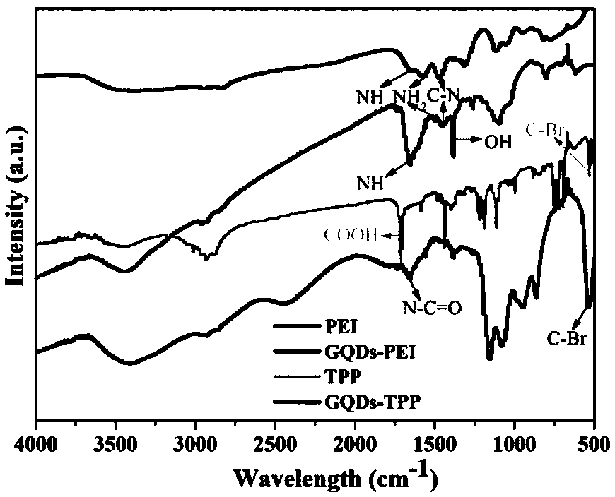
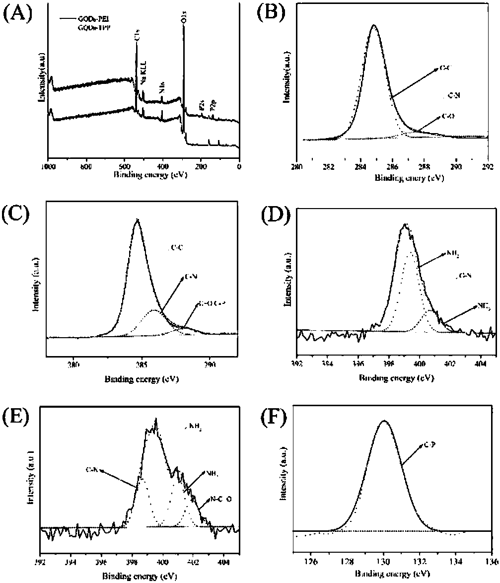
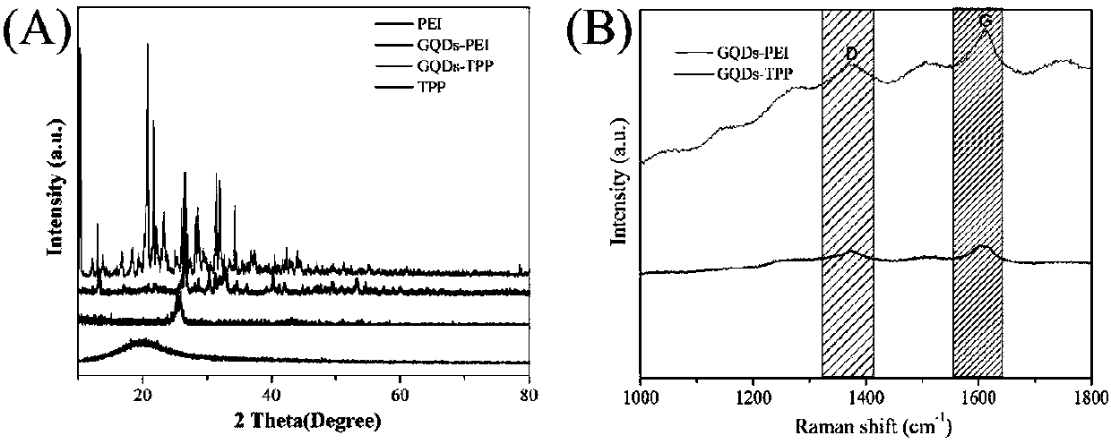

Image

  • Research on preparation and application of graphene quantum dots used for nuclear imaging and mitochondrial imaging
  • Research on preparation and application of graphene quantum dots used for nuclear imaging and mitochondrial imaging
  • Research on preparation and application of graphene quantum dots used for nuclear imaging and mitochondrial imaging

Examples


Embodiment 1

[0065] Provided is a method for preparing graphene quantum dots specifically for targeted imaging of cell nuclei, comprising the following steps:

[0066] 1) 0.1g PEI and 40mL 5mg / mL 1,3,6-trinitropyrene were sonicated for 3-5h and mixed evenly. The mixed solution was transferred to a reactor, and reacted at 180° C. for 10 h. After the reaction was completed, the reactor was naturally cooled to room temperature.

[0067] 2) Filter the reaction solution obtained in step 1) through a 0.22 μm microporous membrane to remove larger-sized carbon particles and insoluble matter.

[0068] 3) The filtrate was placed in a dialysis bag with a molecular weight of 1000 for dialysis, and the water was changed every 6 hours for 3 days.

[0069] 4) Freeze-drying the GQDs-PEI solution prepared in step 3) to obtain GQDs-PEI powder.

[0070] Provided is a method for preparing graphene quantum dots for specific imaging of mitochondria, comprising the following steps:

[0071] 1) Mix 0.1g TPP wi...

Embodiment 2

[0078] Provided is a method for preparing graphene quantum dots for specific targeted imaging of cell nuclei, comprising the following steps:

[0079] 1) 0.1g PEI and 40mL 5mg / mL 1,3,6-trinitropyrene were sonicated for 3-5h and mixed evenly. The mixed solution was transferred to a reactor, and reacted at 200°C for 10 h. After the reaction was completed, the reactor was naturally cooled to room temperature.

[0080] 2) Filter the reaction solution obtained in step 1) through a 0.22 μm microporous membrane to remove larger-sized carbon particles and insoluble matter.

[0081] 3) The filtrate was placed in a dialysis bag with a molecular weight of 1000 for dialysis, and the water was changed every 6 hours for 3 days.

[0082] 4) Freeze-drying the GQDs-PEI solution prepared in step 3) to obtain GQDs-PEI powder.

[0083] Provided is a method for preparing graphene quantum dots for specific imaging of mitochondria, comprising the following steps:

[0084] 1) Mix 0.1g TPP with 100...

Embodiment 3

[0091] Provided is a method for preparing graphene quantum dots for specific targeted imaging of cell nuclei, comprising the following steps:

[0092] 1) 0.1g PEI and 40mL 5mg / mL 1,3,6-trinitropyrene were ultrasonically mixed for 3-5 hours. The mixed solution was transferred to a reactor and reacted at 200°C for 20 hours. After the reaction was completed, the reactor was naturally cooled to room temperature.

[0093] 2) Filter the reaction solution obtained in step 1) through a 0.22 μm microporous membrane to remove larger-sized carbon particles and insoluble matter.

[0094] 3) The filtrate was placed in a dialysis bag with a molecular weight of 1000 for dialysis, and the water was changed every 6 hours for 3 days.

[0095] 4) Freeze-drying the GQDs-PEI solution prepared in step 3) to obtain GQDs-PEI powder.

[0096] Provided is a method for preparing graphene quantum dots for specific imaging of mitochondria, comprising the following steps:

[0097] 1) Mix 0.1g TPP with 1...


PUM

PropertyMeasurementUnit
Particle sizeaaaaaaaaaa

Abstract

The invention discloses graphene quantum dots used for targeting nuclear imaging and mitochondrial imaging. According to the invention, polyethyleneimine (PEI) and 1,3,6-trinitropyrene are subjected to a reaction under hydrothermal reaction conditions so as to form PEI-modified graphene quantum dots (GQDs-PEI) used for nuclear targeting imaging. Based on the constructed GQDs-PEI, 4-carboxybutyltriphenylphosphonium bromide (TPP) is grafted onto the surfaces of GQDs-PEI by an amidation reaction so as to construct GQDs-TPP used for mitochondrial targeting imaging. The invention provides preparation methods and application of GQDs-PEI and GQDs-TPP. The invention has the following beneficial effects: nanotechnology is used for preparing the GQDs-PEI and the GQDs-TPP which are respectively usedfor nuclear targeting imaging and mitochondrial targeting imaging; the preparation methods are simple in process, free of special equipment requirements, friendly to environment, low in cost and capable of realizing large-scale preparation; and the GQDs-PEI and GQDs-TPP of the invention have good biocompatibility and optical properties, and can realize specific targeting imaging of cell nucleusesand mitochondria.

Description

technical field [0001] The invention relates to the technical field of biomedical materials, in particular to a graphene quantum dot for cell nucleus imaging and mitochondrial imaging, a preparation method and application thereof. Background technique [0002] Graphene quantum dots have the characteristics of strong fluorescence brightness, good biocompatibility, high water dispersion and low photobleaching, which make them superior to traditional fluorescent dyes and inorganic quantum dots in optical imaging, optical Sensing, photovoltaic cells and light-emitting diodes have been widely used. [0003] In terms of fluorescence imaging, graphene quantum dots prepared by various synthesis methods are mainly used for imaging various types of cells, including tumor cells, nerve cells, and stem cells. Since most of the currently prepared graphene quantum dots do not have target selectivity, they can only image the cytoplasm, and there is a lack of relevant research on nuclear im...

Claims


Application Information

Patent Timeline
IPC IPC(8): C01B32/184B82Y40/00G01N21/64
CPCB82Y40/00C01P2002/72C01P2002/82C01P2004/01C01P2004/04G01N21/6428
Inventor 范增杰
Owner LANZHOU UNIVERSITY
Why Eureka
Try Eureka