Application of human histone h3 Ser10 and Ser28 in identification of developmental staging of human early embryo
A technology of h3ser10 and histones, applied in analytical materials, instruments, measuring devices, etc., can solve the problems of different phosphorylation patterns and no report on the phosphorylation status of histone H3, and achieve the effect of improving accuracy
- Summary
- Abstract
- Description
- Claims
- Application Information
AI Technical Summary
Problems solved by technology
Method used
Image
Examples
Embodiment 1
[0032] Embodiment 1, material and method
[0033] 1. Research object
[0034] The subjects of this study were patients who underwent IVF treatment in the Reproductive Center of the First Affiliated Hospital of Zhengzhou University. Inclusion criteria: 1. Age ≤ 35 years old; 2. Follicle stimulating hormone (FSH) < 12mIU / L; 3. The row stimulation program is a long program or an ultra-long program. All patients had signed the "Informed Consent Form", and the study was approved by the Medical Ethics Committee of Zhengzhou University.
[0035] 2. Obtaining oocytes
[0036] The collected oocytes were immature oocytes during intracytoplasmic sperm injection (ICSI). Conventionally, oocytes are retrieved 36 hours after injection of human chorionic gonadotropin (Hcg), and intracytoplasmic sperm injection is performed 4-6 hours after oocyte retrieval. At this time, only oocytes that have grown to the metaphase of the second meiosis are considered mature oocytes and can be injected. ...
Embodiment 2
[0053] Example 2, Localization of H3Ser10 and H3Ser28 Phosphorylation in Human Oocytes
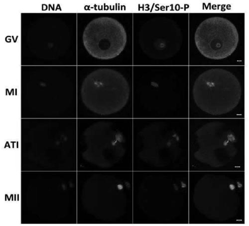
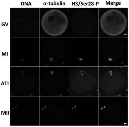
[0054] A total of 104 oocytes were collected, including 28 at the GV stage, 26 at the MI stage, 24 at the ATI stage, and 26 at the MII stage. Immunofluorescence staining analysis of cloned antibodies and monoclonal antibodies against histone H3 phosphorylation at serine 28.
[0055] Such as figure 1 As shown, the results of immunofluorescence analysis of monoclonal antibody against histone H3 serine phosphorylation at position 10 showed that phosphorylation of H3 Ser10 was always present during oocyte maturation (see figure 1 Column H3 / Ser10-P in ), and start in GV stage oocytes, and co-localize with DNA, and have been distributed on chromosomes.
[0056] Such as figure 2 As shown, the results of immunofluorescence analysis of anti-histone H3 phosphorylation serine 28 monoclonal antibody showed that during oocyte maturation, the phosphorylation signal of H3Ser28 was not found in the GV...
Embodiment 3
[0057] Example 3, Localization of phosphorylation of H3Ser10 and H3Ser28 in 3PN early embryos derived from IVF
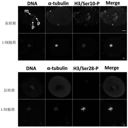
[0058] A total of 85 3PN zygotes were collected and divided equally into H3Ser10 and H3Ser28 groups. Immunofluorescence staining analysis was performed with anti-histone H3 serine 10 phosphorylated monoclonal antibody and anti-histone H3 serine 28 phosphorylated monoclonal antibody respectively.
[0059] The result is as image 3Shown: Immunofluorescence staining revealed that in human 3PN fertilized eggs, the localization of phosphorylation of H3Ser10 and H3Ser28 was different. H3Ser10 phosphorylation mainly appeared in the male pronucleus at the 3PN pronuclear stage, and was evenly distributed at the 1-cell stage with further development. on the chromosome. For H3Ser28, there was no phosphorylation signal at the 3PN prokaryotic stage, but with the further development of the embryo, it could be seen that it was located on the chromosome at the 1-cell stage.
PUM
 Abstract
Description
Claims
Application Information
 - R&D
- Intellectual Property
- Life Sciences
- Materials
- Tech Scout
- Unparalleled Data Quality
- Higher Quality Content
- 60% Fewer Hallucinations
Browse by: Latest US Patents, China's latest patents, Technical Efficacy Thesaurus, Application Domain, Technology Topic, Popular Technical Reports.
© 2025 PatSnap. All rights reserved.Legal|Privacy policy|Modern Slavery Act Transparency Statement|Sitemap|About US| Contact US: help@patsnap.com


