Method for discriminating liver cancer differentiation grades by using multiphoton imaging technology

A multi-photon imaging and multi-photon technology, applied in the preparation of test samples, material excitation analysis, etc., can solve time-consuming problems and achieve the effect of overcoming time-consuming

Inactive Publication Date: 2018-08-21
FUJIAN NORMAL UNIV
View PDF0 Cites 2 Cited by
  • Summary
  • Abstract
  • Description
  • Claims
  • Application Information

AI Technical Summary

Problems solved by technology

The liver cancer differentiation grade data obtained by this label-free rapid discrimination method is compared with the pathologist's diagnosis data to achieve the purpose of objectively evaluating the tumor differentiation grade and effectively overcome the shortcomings of time-consuming, missed diagnosis and strong subjectivity in current clinical biopsy techniques

Method used


Image

  • Method for discriminating liver cancer differentiation grades by using multiphoton imaging technology
  • Method for discriminating liver cancer differentiation grades by using multiphoton imaging technology

Examples


Embodiment 1

[0025] (1) Sampling

[0026] 200 cases of diseased liver cancer tissues were removed from patients by cutting, forceps or puncture, that is, fresh specimens without fixation.

[0027] (2) Quick-frozen film production

[0028] The sample is quickly frozen in the cryostat to below minus 18°C ​​and then sliced. When slicing, the rocker handle is pushed back and forth rhythmically, and the slice is complete and thin (thickness 8-9 μm), and then it can be attached to the glass slide. The cut slices were fixed with 95% ethanol and sealed.

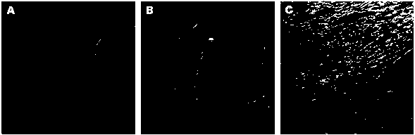
[0029] (3) Multiphoton imaging of collagen fibers inside the tumor

[0030] Using a laser scanning microscopy imaging system, set the excitation wavelength to 810 nm, and select an upright 10× objective lens (0.45 NA) to scan the sample. Set up 2 receiver channels, one at 370-419 nm, marked green, for collecting SHG signals, and the other 420-700 nm, marked red, for detecting TPEF signals. Single image size is 8×8 mm 2 , 512×512 pixel, a typic...


PUM

PropertyMeasurementUnit
wavelengthaaaaaaaaaa

Abstract

The invention relates to a method for discriminating liver cancer differentiation grades by using a multiphoton imaging technology. The method comprises the following steps: firstly, carrying out quick freezing film shooting processing on a fresh liver cancer specimen subjected to surgical excision or puncture sampling; secondly, obtaining a collagen fiber MPM image inside a tumor by multiphoton microscopic imaging, processing the image, extracting features and then carrying out statistical analysis and discrimination by using Mann-Whitney inspection and Boxplot. According to the method disclosed by the invention, the multiphoton microscopic imaging and an image processing algorithm are utilized to be combined with a statistical analysis method for evaluating different differentiation grades of liver cancer; liver cancer differentiation grade data obtained by a label-free quick discrimination method are compared with pathologist diagnostic result data so as to achieve the aim of objectively evaluating the liver cancer differentiation grades; and the defects of time consumption, missed diagnosis and high subjectivity of a current clinical biopsy technique are effectively overcome.

Description

technical field [0001] The invention belongs to the field of biomedicine, and relates to a method for discriminating the differentiation grade of liver cancer by using multiphoton imaging technology. Specifically, fresh specimens of liver cancer taken by surgery or puncture are subjected to frozen section processing, and the collagen fibers inside the tumor are imaged by a multiphoton microscope. For the second harmonic generation (SHG) signal, Mann-Whitney test and Boxplot box plot are used for statistical analysis and discrimination. Background technique [0002] Primary liver cancer is the fifth most common cancer in the world and the third most deadly cancer. According to cancer statistics in China in 2015, there were an estimated 466,100 new cases and 422,100 deaths, among which the male and female mortality rates were the third and fourth in cancer mortality, respectively. More than 90% of primary liver cancer is hepatocellular carcinoma, which is caused by chronic li...

Claims


Application Information

Patent Timeline
Patent Type & Authority Applications(China)
IPC IPC(8): G01N21/63G01N1/42
CPCG01N21/63G01N1/42
Inventor 林宏心王光兴陈瑚左宁卓双木
Owner FUJIAN NORMAL UNIV
Why Eureka
Try Eureka