Cancer detection system based on exosome and method thereof
A cancer detection and exosome technology, applied in the field of exosome-based cancer detection systems, can solve problems such as lack of analysis methods, and achieve the effect of promoting accumulation and convenient operation
- Summary
- Abstract
- Description
- Claims
- Application Information
AI Technical Summary
Problems solved by technology
Method used
Image
Examples
Embodiment 1
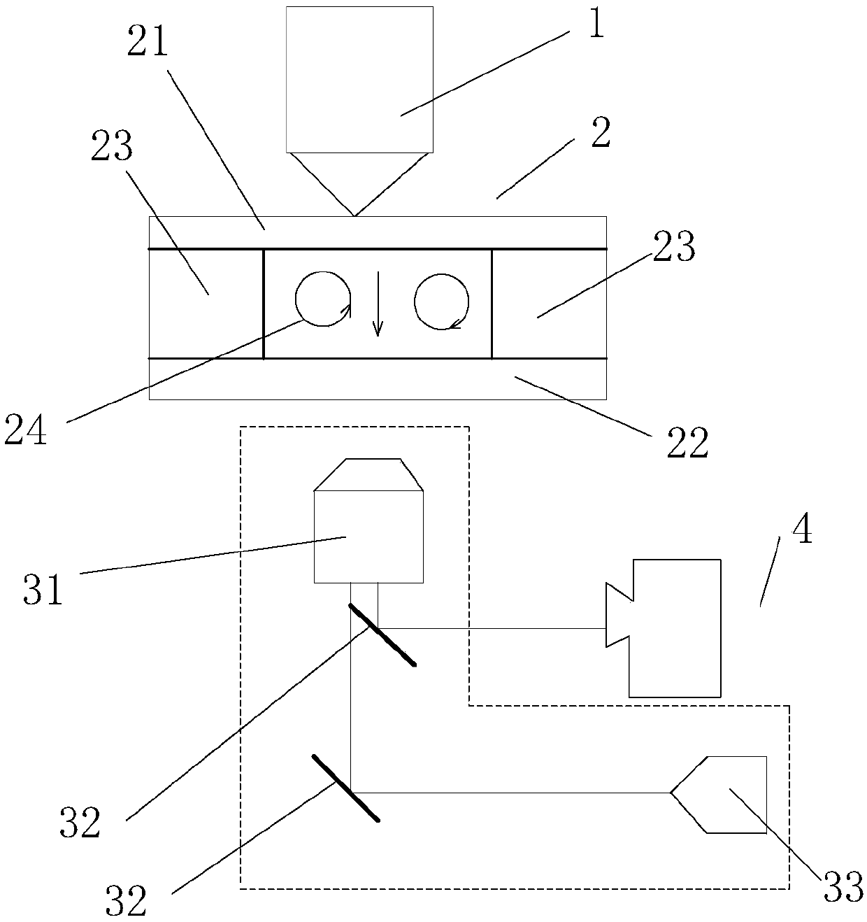
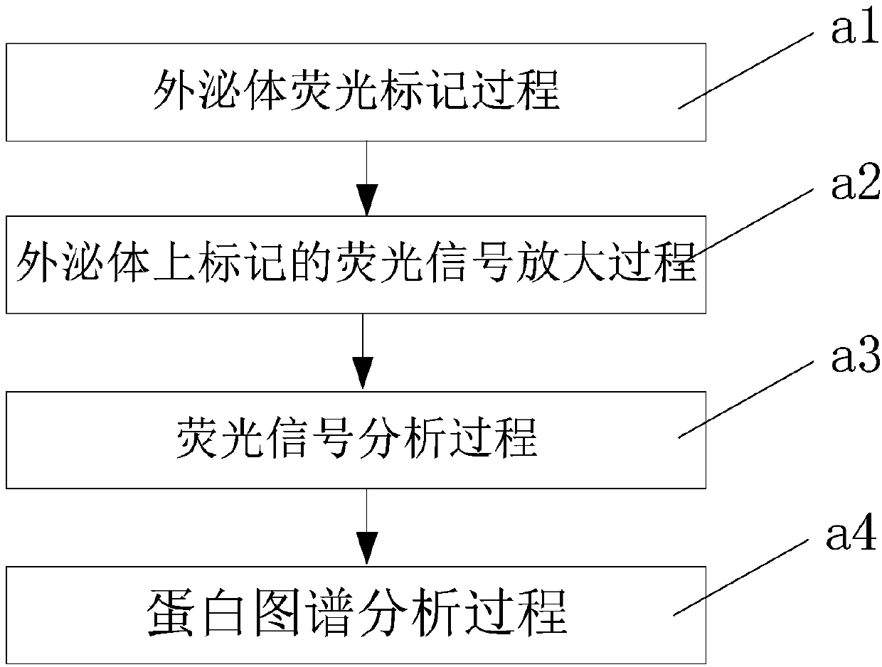
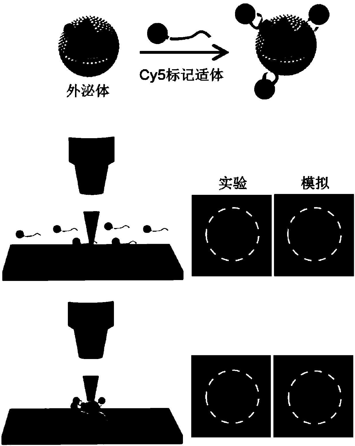
[0082] Incubate exosome samples with fluorescently labeled aptamers, and the selected aptamers are oligonucleotides that can specifically bind to proteins or other small molecular substances screened by the in vitro screening technique SELEX (Systematic Evolution of Ligands by Exponential Enrichment) Fragments, specifically, the fluorescently labeled aptamer is a 40-base single-stranded DNA, and the diameter of the coil in the sample liquid is less than 5 nanometers, while the diameter of the exosome is 30-150 nanometers; it will specifically recognize the CD63 protein Aptamers were applied to exosomes in the culture supernatant of A375 cells (human melanoma cells). Fluorescent groups can be modified at the end of the aptamer by standard means. When the aptamer specifically interacts with the target protein on the surface of the exosome, the exosome is labeled with the fluorescence carried by the aptamer. The exosome sample in this example is the supernatant of the cell cultur...
Embodiment 2
[0087] In this example, using serum samples from patients with cervical cancer, 7 different aptamers were used to detect the abundance of 7 surface proteins (CD63, PTK7, EpCAM, HepG2, HER2, PSA, CA125) of exosomes in serum samples, and Comparison with healthy human serum samples.
[0088] The exosome manipulation method used, as well as the laser, sample chamber, microscope and CCD camera are the same.
[0089] combine Figure 4 As shown, it can be seen that the serum exosomes of this cervical cancer patient highly express CD63 protein, and cancer-related markers PTK7, EpCAM, HepG2, HER2, PSA and CA125, among which CA125 can be used as a traditional cervical cancer marker, and some cervical cancer patients There is high expression of HER2. It is generally believed that tumor markers PTK7 and EpCAM are related to various cancers, HepG2 is mainly specific to liver cancer, and PSA is mainly specific to prostate cancer. However, these tumor markers are not strictly correlated w...
Embodiment 3
[0093] In this embodiment, the micro-nano particles used are non-biological micro-nano particles, specifically fluorescent polystyrene microspheres, the brand is Thermofisher, the diameter is 50 to 200 nanometers, the mass fraction is 0.001%, and it is dissolved in Tween20 containing 0.02%. in aqueous solution. The laser, the sample chamber, the microscope and the CCD camera are all the same as the above-mentioned embodiments 1 and 2.
[0094] combined Figure 5 As shown, all the fluorescent microspheres with different diameters are highly converged at the laser spot, and according to the gray value of the fluorescence measurement and the fluorescence picture, it can be seen that the degree of convergence and the fluorescence intensity increase with the increase of the particle diameter, which is consistent with the work of this embodiment The principle is consistent, that is, large particles tend to aggregate more. This example illustrates that both biological and non-biolo...
PUM
| Property | Measurement | Unit |
|---|---|---|
| Thickness | aaaaa | aaaaa |
Abstract
Description
Claims
Application Information
 - R&D
- Intellectual Property
- Life Sciences
- Materials
- Tech Scout
- Unparalleled Data Quality
- Higher Quality Content
- 60% Fewer Hallucinations
Browse by: Latest US Patents, China's latest patents, Technical Efficacy Thesaurus, Application Domain, Technology Topic, Popular Technical Reports.
© 2025 PatSnap. All rights reserved.Legal|Privacy policy|Modern Slavery Act Transparency Statement|Sitemap|About US| Contact US: help@patsnap.com


