Detection method of ranavirus in water

A detection method, frog virus technology, applied in the directions of microorganism-based methods, biochemical equipment and methods, and microbial determination/inspection, etc., to avoid false negatives and to be easily identified.

Inactive Publication Date: 2018-11-02
SICHUAN AGRI UNIV
View PDF6 Cites 3 Cited by
  • Summary
  • Abstract
  • Description
  • Claims
  • Application Information

AI Technical Summary

Problems solved by technology

However, there is no method for dete

Method used


Image

  • Detection method of ranavirus in water
  • Detection method of ranavirus in water
  • Detection method of ranavirus in water

Examples


Embodiment 1

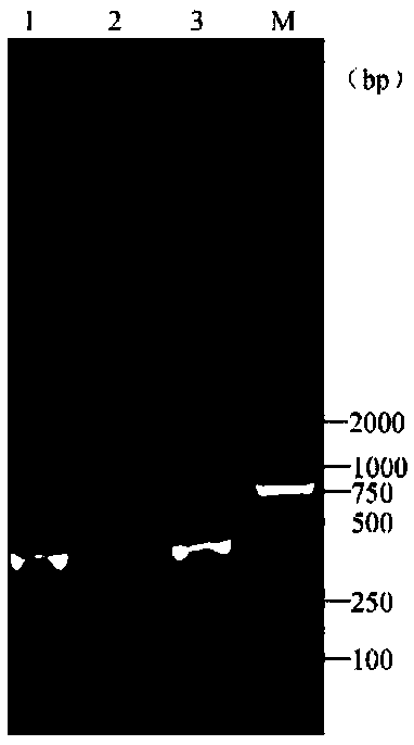
[0060] Water sample virus enrichment:

[0061] Take 1L of aquaculture water sample and use medical gauze to filter out impurities, and use 5mol / L HCl to adjust the pH of the filtered water sample to 3.5. Add 10gNaCl and 24gAlCl to the adjusted pH water sample 3 ·6H 2 O to stand for 5 hours. After standing still, a flocculent precipitate will be formed at the bottom. Pour off part of the supernatant. Use a 0.45 μL filter membrane to filter out the excess water at the bottom of the flocculent precipitate. Dry the filtered flocculent precipitate and put it into an EP tube. , add 1mL of PBS buffer solution and pipette to dissolve, and the obtained solution is the solution after the water body virus is enriched.

[0062] DNA extraction:

[0063] After the virus-enriched solution was repeatedly frozen and thawed three times, 1 mL was drawn into a test tube, and centrifuged at 13,000 r / min for 5 min at a temperature of 4°C. Take 400 μL of centrifuged supernatant, add 500 μL of sa...

Embodiment 2

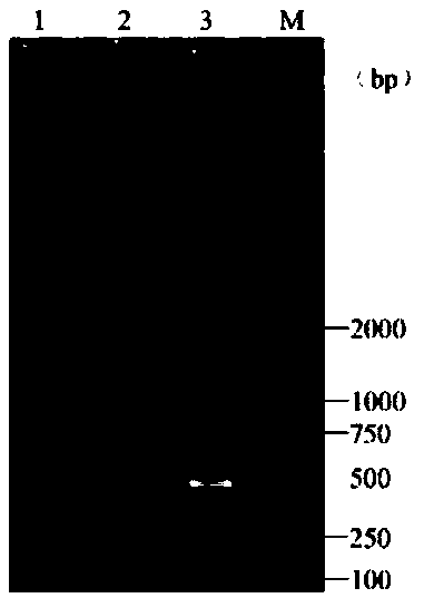
[0077] Take 1L of tap water, add 2mL of TCID to the tap water 50 The frog virus cell culture fluid of 10.648 is used as the aquaculture water sample to be tested, and the steps in Example 1 are used to successively carry out water sample virus enrichment, DNA extraction and PCR detection, and finally use the product after the PCR reaction after agarose gel electrophoresis The gel imager takes pictures such as figure 2 As shown, a main band appears between maker 250bp-500bp in lane 1, which is judged to be positive for RAV, indicating that frog virus was detected. Then take 1L of tap water without adding any reagents and follow the steps in Example 1 to carry out water sample virus enrichment, DNA extraction and PCR detection in sequence, and finally the product after the PCR reaction is subjected to agarose gel electrophoresis and then photographed with a gel imager as shown figure 2 No bands were added in the 2 swimming lanes shown, and it was judged to be RAV negative, wh...

Embodiment 3

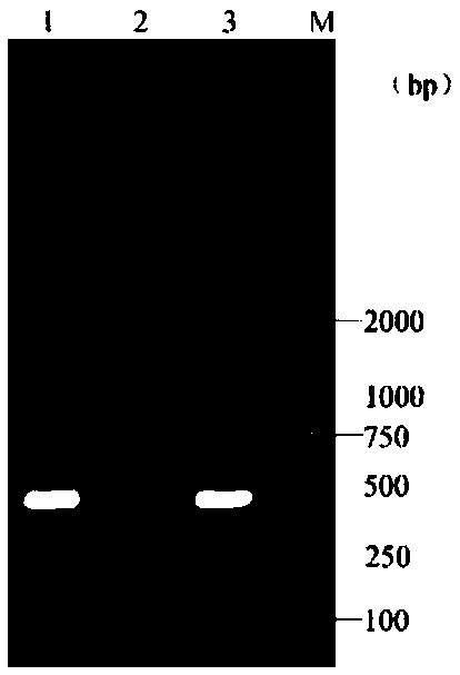
[0079] Take 1L of well water and add 2mL of TCID to the well water 50 The frog virus cell culture fluid of 10.648 is used as the aquaculture water sample to be tested, and the steps in Example 1 are used to successively carry out water sample virus enrichment, DNA extraction and PCR detection, and finally use the product after the PCR reaction after agarose gel electrophoresis The gel imager takes pictures such as image 3 A main band appears between maker250bp-500bp in lane 1 shown, which is judged to be positive for RAV, indicating that frog virus was detected. Then take 1L of well water without adding any reagents and follow the steps in Example 1 to carry out water sample virus enrichment, DNA extraction and PCR detection in sequence, and finally perform agarose gel electrophoresis on the products after the PCR reaction, and use a gel imager to take pictures as follows: image 3 No bands were added in the 2 lanes shown, and it was judged to be negative for RAV, indicating...


PUM


Abstract

The invention relates to a detection method of ranavirus in water and belongs to the field of virus detection. The detection method of the ranavirus in culture water is constructed in combination witha method for enriching viruses in water and a PCR technology. A method for enriching viruses in water with a NaCl-AlCl3.6H2O precipitation method is improved, and the enrichment time is shortened. Specific PCR primers are improved, multiple sequence alignment is performed on MCP (major capsid protein) of ranavirus including giant salamander ranavirus, rana tigrina ranavirus, common midwife toad virus and type-3 ranavirus, primers are designed for conservative areas of sequences, so that false negative of detection due to sequence variation of viruses in the evolutionary process is avoided, the blank of methods for detecting ranavirus in water on the market at present is effectively filled up, and the method has great social benefits and economic benefits.

Description

technical field [0001] The invention relates to the field of virus detection, in particular to a method for detecting frog virus in water. Background technique [0002] Ranavirus is a DNA virus that harms fish, amphibians, and reptiles. It can infect a variety of aquatic economically farmed animals such as Chinese soft-shelled turtles, tiger frogs, black-spotted frogs, giant salamanders, and snapping turtles. , causing huge economic losses to the aquaculture industry. The pathogen can spread infection horizontally through water, so rapid detection of frog virus in aquaculture water is particularly important to guide disease prevention and control strategies. However, there is no method for detecting frog virus in aquaculture water. Contents of the invention [0003] In order to solve the above-mentioned technical problems, the present invention provides a method for detecting frog virus in water bodies, which fills the gap in the field of current detection of frog virus ...

Claims


Application Information

Patent Timeline
IPC IPC(8): C12Q1/70C12Q1/686C12R1/93
CPCC12Q1/686C12Q1/701C12Q2565/125
Inventor 耿毅余泽辉牟维豪王世震白明焕欧阳萍陈德芳黄小丽
Owner SICHUAN AGRI UNIV
Why Eureka
Try Eureka