Fluorescent quantitative PCR primer for detecting mycoplasma, detection method and application

A fluorescent quantitative and mycoplasma technology, applied in the field of pathogen detection, achieves the effect of good repeatability, high sensitivity, and rapid repeatability detection

Inactive Publication Date: 2018-11-27
安徽古一生物科技有限公司
View PDF4 Cites 2 Cited by
  • Summary
  • Abstract
  • Description
  • Claims
  • Application Information

AI Technical Summary

Problems solved by technology

[0005] The technical problem solved by the present invention is that the prior art cannot quickly and quantitatively diagnose the contamination of mycoplasma in the cell culture fluid

Method used


Image

  • Fluorescent quantitative PCR primer for detecting mycoplasma, detection method and application
  • Fluorescent quantitative PCR primer for detecting mycoplasma, detection method and application
  • Fluorescent quantitative PCR primer for detecting mycoplasma, detection method and application

Examples


Embodiment 1

[0031] Design and synthesis of real-time fluorescent quantitative PCR primers for quantitative detection of mycoplasma in cell culture medium

[0032] (1) Primer design

[0033] Mycoplasma (M.orale), Mycoplasma fermentans (M.Fermentans), Mycoplasma arginini (M.arginini), Mycoplasma inflammatoryis (M. Arthritidis), Mycoplasma hyorhinis (M.hyorhinis) and A.laidlawii (A.laidlawii) 16S rRNA gene sequences were compared with DNA Man software, and according to the principle of fluorescent quantitative PCR primer design, Primerpremier5. 0, for the 16S rRNA gene of mycoplasma (this is the conserved region of mycoplasma), design upstream and downstream primers, primers are PAGE purification, and the described fluorescent quantitative PCR primer sequence that is used to detect mycoplasma is as follows:

[0034] Upstream primer: 5'-ACTCCTACGGGAGGCAGCAGTA-3' (as shown in SEQ NO.1)

[0035] Downstream primer: 5'-GATTACTAGCGATTCCGACTTCAT-3' (as shown in SEQ NO.2)

[0036] The synthesis o...

Embodiment 2

[0038] Real-time Fluorescent Quantitative PCR Detection of Mycoplasma in Cell Culture with Fluorescent Quantitative PCR Primers

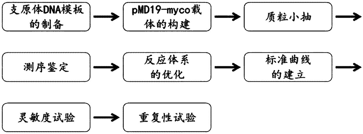
[0039] figure 1 It is a technical roadmap of the real-time fluorescent quantitative PCR detection method for mycoplasma in cell culture solution of the present invention;

[0040] (1) Preparation of samples to be tested

[0041] a. Transfer 100 μL of cell culture medium to a 1.5 mL sterile EP tube, make sure that the EP tube cap is tightly sealed to prevent evaporation;

[0042] Adherent cells: After 2-3 days of cell culture, grow to a confluence of 80%-90%; Suspension cells: After 2-3 days of cell culture, the cell density reaches 5×10 5 -1×10 6 about;

[0043] b. Transfer 50 μL of cell culture solution to a sterile PCR tube, and heat-treat at 95°C for 5 minutes;

[0044] c. 12000rpm for 5s, take the supernatant and transfer it to a sterile PCR tube, and the sample can be detected immediately, or stored in a -20 degree refrigerator for frozen ...

Embodiment 3

[0060] Detection of mycoplasma samples in cell culture medium

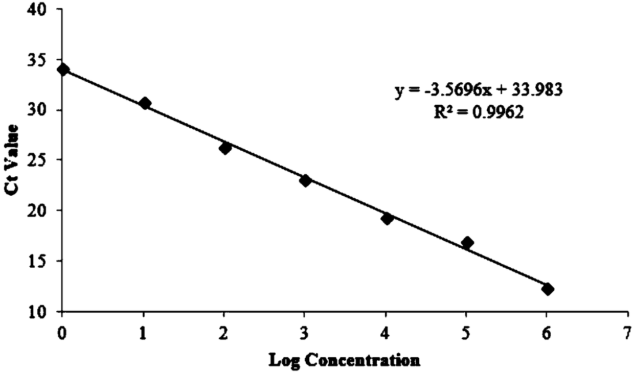
[0061] Collect 10 samples of positive contamination of cell culture medium, and process the samples: transfer 100 μL of cell culture medium to a 1.5 mL sterile EP tube, and ensure that the cap of the EP tube is tightly sealed to prevent evaporation; take 50 ul of cell culture medium and transfer it to a sterile PCR tube Heat treatment at 95°C for 5min; 12000rpm for 5s, transfer the supernatant to a sterile PCR tube. Three replicate wells were set up for each group of samples, and a negative control (adding fresh medium, NTC) was set, and the real-time fluorescent quantitative PCR detection of mycoplasma in the cell culture medium was carried out with the reaction system and fluorescent quantitative PCR reaction conditions in step 3. The test results are: all 10 cell culture fluids are positive for mycoplasma contamination, and the CT value of the sample is substituted into the linear regression equation of the sta...


PUM


Abstract

The invention discloses a fluorescent quantitative PCR primer for detecting mycoplasma, a detection method and application, relates to the field of pathogen detection and aims to solve the problem that the prior art cannot quickly and quantitatively diagnose contamination of mycoplasma in cell culture liquid. The invention further discloses a kit. The fluorescent quantitative PCR primer has the advantages of high sensitivity, excellent repeatability, quickness in detection, little pollution and low cost and can be used for quantitatively identifying mycoplasma in the cell culture liquid in scientific research, production and clinical experiments.

Description

technical field [0001] The invention relates to the field of pathogen detection, in particular to a fluorescent quantitative PCR primer for detecting mycoplasma, a detection method and an application. Background technique [0002] Mycoplasma is the smallest prokaryotic cell-type microorganism that has no cell wall and is mostly irregular spherical and filamentous. It can pass through a 0.22 μm bacterial filter and can grow and reproduce in an inanimate medium. In the process of cell culture, the cells are easily polluted by mycoplasma. Domestic and foreign studies have shown that more than 98% of the cells are polluted by mycoplasma in the following six types: M.orale, M.Fermentans, M.arginini. .arginini), M. arthritidis, M. hyorhinis and A. laidlawii. [0003] Due to their tiny size (~100 nm), mycoplasma cannot be detected with the naked eye or light microscope, and therefore they usually go undetected for a long time. Furthermore, due to their lack of a cell wall, they a...

Claims


Application Information

Patent Timeline
Patent Type & Authority Applications(China)
IPC IPC(8): C12Q1/689C12Q1/686C12Q1/04C12N15/11C12R1/35
CPCC12Q1/686C12Q1/689C12Q2563/107C12Q2545/114
Inventor 李杰刘振云王维程丰伟徐华栋
Owner 安徽古一生物科技有限公司
Why Eureka
Try Eureka