Preparation and cryopreservation method and application of human placental subchorionic large blood vessel tissue

A cryopreservation method and chorionic membrane technology, applied in the preservation, application, animal husbandry and other directions of human or animal body, can solve the problems of the influence of cell activity, loss of the function of cryopreserved tissue to protect cells, etc., and achieve the effect of improving activity

Active Publication Date: 2021-10-01
JIANGXI YINFENG DINGCHENG BIO ENG
View PDF5 Cites 0 Cited by
  • Summary
  • Abstract
  • Description
  • Claims
  • Application Information

AI Technical Summary

Problems solved by technology

Therefore, if the volume of cryopreserved tissue is too small, the cells will be more exposed to the toxic components of the cryoprotectant, the cell viability will be affected, and the function of cryopreserved tissue to protect cells will be lost.

Method used


Image

  • Preparation and cryopreservation method and application of human placental subchorionic large blood vessel tissue
  • Preparation and cryopreservation method and application of human placental subchorionic large blood vessel tissue
  • Preparation and cryopreservation method and application of human placental subchorionic large blood vessel tissue

Examples


Embodiment 1

[0038] The cryopreservation method of human placental subchorionic large blood vessel tissue of embodiment 1

[0039] Placenta collection: Select a healthy placenta without infectious diseases and obstetric complications, obtain the consent of the parturient and sign the informed consent; for normal collection, the collected placenta will be transported to the laboratory within 48 hours, and various necessary tests will be carried out, such as Detection of infectious diseases such as viruses, detection of bacterial contamination, etc.

[0040] The method of cryopreservation, the steps are as follows:

[0041] (1) Wash the placenta (to remove dirt and microbial contamination); cut off the decidua along the edge of the placenta, and remove the amniotic membrane.

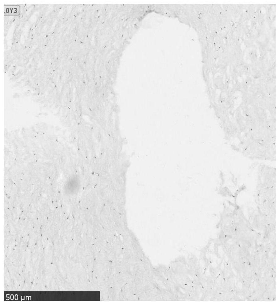
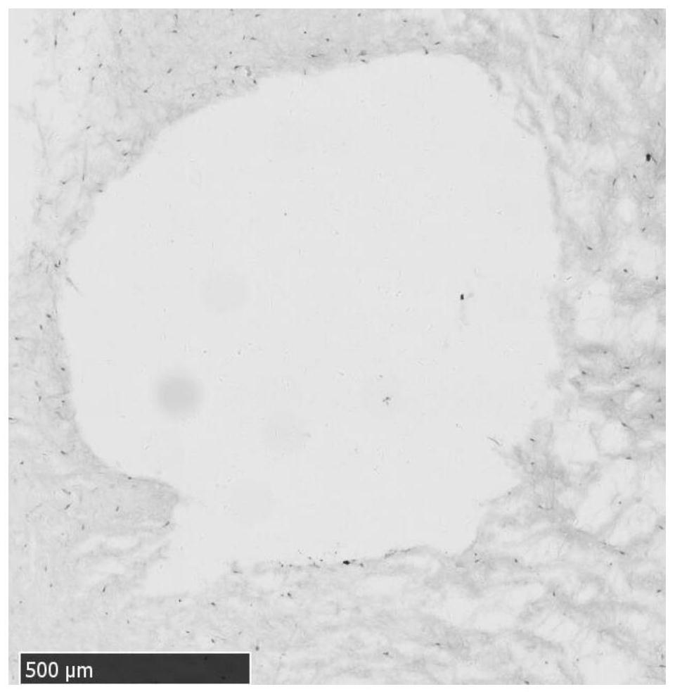
[0042] (2) Separate the above-mentioned placental fetal surface chorion and large blood vessels from which the amniotic membrane has been removed (keep the subchorionic large blood vessels intact as much as possible),...

Embodiment 2

[0050] Recovery after embodiment 2 cryopreservation

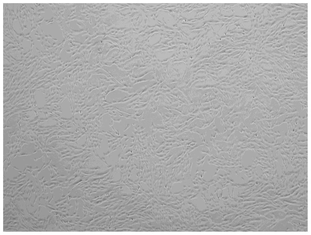
[0051] According to the method of Example 1, after cryopreservation for 6 months, recovery was carried out, and mesenchymal stem cells were induced and isolated. The steps were as follows:

[0052] Take the cryopreserved placental subchorionic large blood vessel tissue out of the liquid nitrogen, place it in the gas phase for 10 minutes to equilibrate, and then quickly place the cryopreservation bag in a water bath at 37°C to 42°C; quickly transfer the cryopreservation bag to a safety cabinet after dissolving , open the cryopreservation bag, gently remove the placental subchorionic large blood vessel tissue with tweezers, put it into the resuscitation solution of three times the reference concentration at 4°C (pre-cooled to 4°C in advance), equilibrate for 1 minute, take it out, and then put it in 4°C ℃ double standard concentration resuscitation solution (pre-cooled to 4 ℃ in advance), equilibrate for 1 to 3 minutes, take ...


PUM


Abstract

The invention discloses a method for preparing and freezing human placental subchorionic macrovascular tissue, comprising: (1) washing the placenta; cutting off the decidua along the edge of the placenta to remove the amniotic membrane; (2) removing the amniotic membrane from the placenta The fetal facial chorion and large blood vessels are separated together, washed with normal saline or PBS buffer, and the blood vessels are flushed to remove blood clots in the blood vessels; then, the large blood vessels connected to the chorionic plate are cut off one by one; (3) the blood vessels are transferred Put it into a cryopreservation tube or a cryopreservation bag, introduce the vitrification solution in a three-step method, transfer it to a programmed cooling device, cool it down to -80°C to -90°C, and transfer it to liquid nitrogen for cryopreservation. The cryopreservation method of the present invention is beneficial to improve the activity of cryopreserved tissues and cells, and the morphology, function, and structure after recovery are consistent with those of fresh tissues. The preserved tissues can not only be used in the fields of separating stem cells and epithelial cells, but also in tissue Engineering transplantation and other fields.

Description

technical field [0001] The invention relates to a method for preparing and freezing human placental subchorionic large blood vessel tissue and its application, in particular to a method for cryopreserving and resuscitating human placental subchorionic large blood vessel tissue. Background technique [0002] The placenta is an important organ for material exchange between the fetus and the mother. It is a tissue-combining organ between the mother and the child formed by the joint growth of the embryonic embryonic membrane and the maternal endometrium during human pregnancy. In recent years, with the deepening of scientific research, people can classify various types of cells from placenta, including hematopoietic stem cells, mesenchymal stem cells, epithelial cells, etc., thus changing the long-term status of placenta as medical waste. [0003] Modern scientific research suggests that placental amniotic membrane can be used not only to expand amniotic membrane-derived mesench...

Claims


Application Information

Patent Timeline
Patent Type & Authority Patents(China)
IPC IPC(8): A01N1/02
CPCA01N1/0221
Inventor 王肇光徐峰波王圣川宋现收生德伟李德柱
Owner JIANGXI YINFENG DINGCHENG BIO ENG
Why Eureka
Try Eureka