Site-specific radioisotope-labeled antibody using IgG-binding peptide
A radioactive and nuclide technology, applied in in vivo radioactive preparations, preparations for in vivo experiments, peptides, etc., can solve the problems of reduced antibody function and high development costs
- Summary
- Abstract
- Description
- Claims
- Application Information
AI Technical Summary
Problems solved by technology
Method used
Image
Examples
Embodiment 1
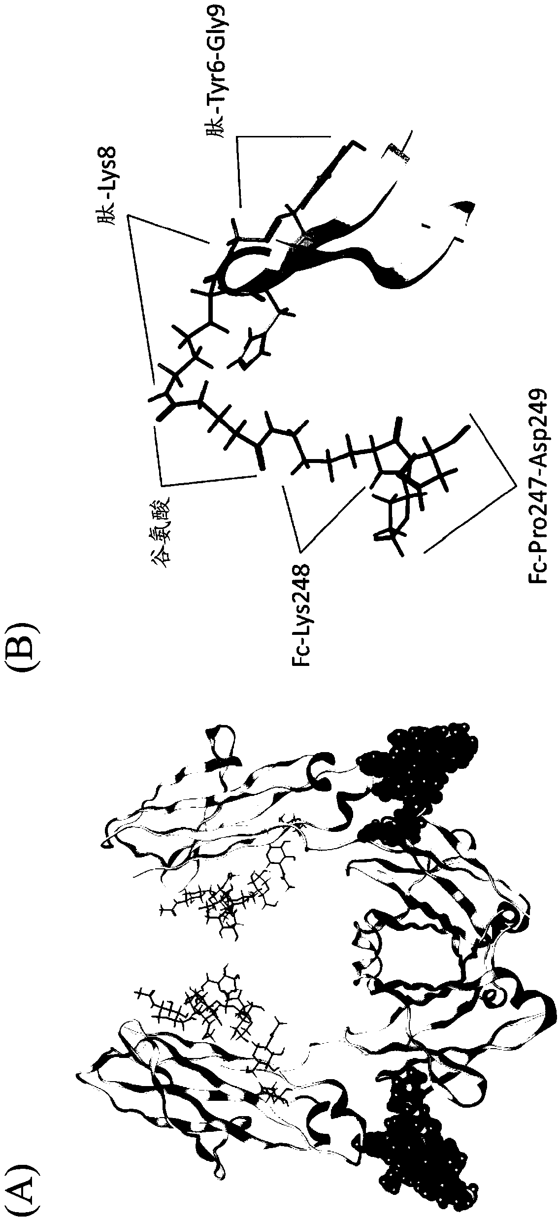
[0310] [Example 1: X-ray crystal structure analysis of the complex of IgG-binding peptide and IgG]
[0311]
[0312] (1) Preparation of IgG binding peptide solution
[0313] By utilizing the peptide solid-phase synthesis method of the F-moc method, a sequence (SEQ ID NO: 31, wherein HC is homocysteine, 4th 2 Cys at position 1 and 14, and 2 homocysteine at position 2 and 16 respectively form a disulfide bond in the molecule) cyclic homocysteine peptide. 0.8 mg of the prepared IgG-binding peptide powder was dissolved in 24 µL of 100% dimethylsulfoxide (Wako Pure Chemical Industries, Ltd.) to prepare an IgG-binding peptide solution.
[0314] (2) Preparation of a complex of Fc and IgG-binding peptide
[0315] In 20 mmol / L phosphate buffer (pH 7.0) containing 10 mM EDTA and 1 mM L-cysteine, the hinge of human IgG (Chugai Pharmaceutical) was digested with papain (manufactured by Roche) at 37 °C. )part. Next, using a cation exchange chromatography column (TSKgel SP5-PW (Tos...
Embodiment 2
[0326] [Example 2: Preparation and properties of peptides for labeling]
[0327]
[0328] Using the Fmoc solid-phase synthesis method and according to conventional methods, synthesize amino-PEG4 modified amino-PEG4 with biotin (Biotin) or 5 / 6-TAMURA succinimidyl ester (AnaSpec company) (fluorescent pigment) The peptide GPDCAYHXGELVWCTFH (SEQ ID NO: 2) was synthesized (where the C-terminus is amidated). After removal of the protecting group, intramolecular S-S bond formation was performed under oxidative conditions in aqueous solution at pH 8.5 using reverse phase HPLC at a flow rate of 1.0 ml / min through a gradient of 10% to 60% acetonitrile containing 0.1% TFA Elution, purification of peptides with intramolecular S-S bonds.
[0329] 100 µL of a DMF solution containing 1 mM of the purified IgG-binding peptide was mixed with 100 µL of an acetonitrile solution of 100 mM DSS or DSG (Thermo Fisher Scientific), and reacted overnight at room temperature. After diluting the react...
Embodiment 3
[0338] [Example 3: Specific modification of human IgG-Fc using IgG-binding peptide]
[0339]
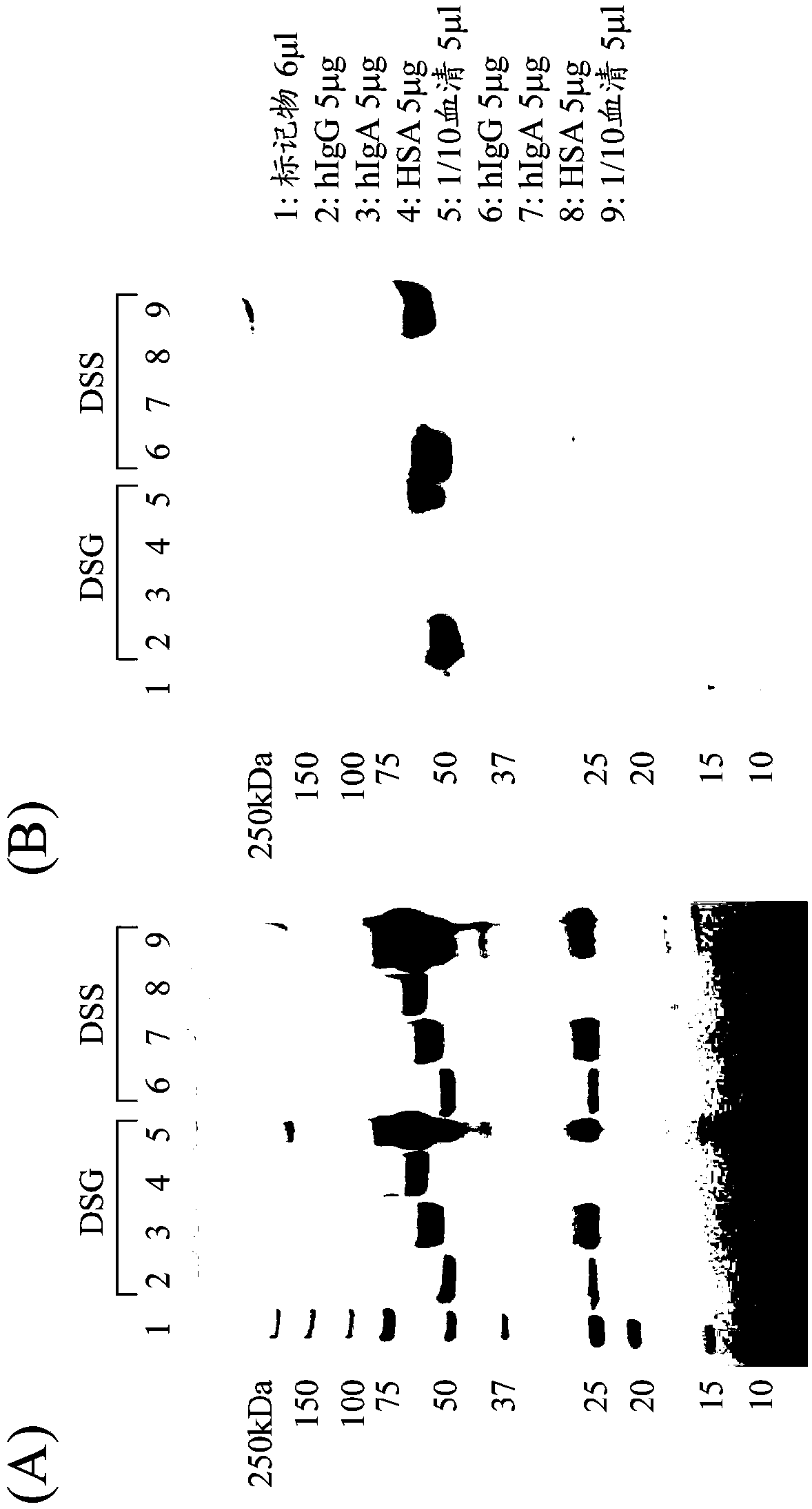
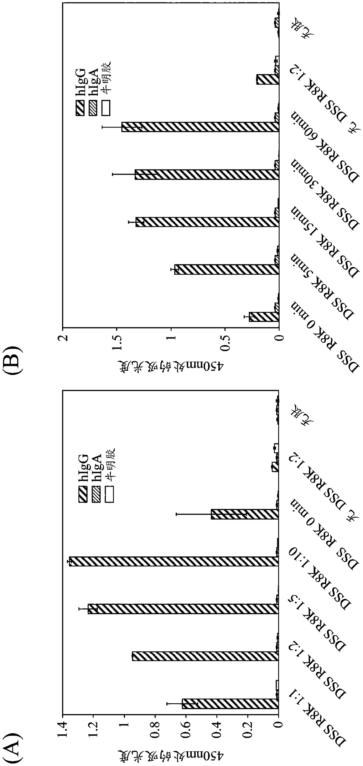
[0340] A labeling reagent peptide modified with DSS or DSG to an IgG-binding peptide (type I (R8K)) with biotin-PEG4 added to the N-terminus was prepared by the same method as in Example 2, and reacted with human IgG Fc to examine human Labeling reaction of IgG Fc. That is, after purifying the IgG-binding peptide (R8K) (200 pmol / 5 μL in 0.1% TFA) reacted with excess DSS or DSG using a reverse-phase column by the same method as in Example 2, acetonitrile was removed under reduced pressure, and then about 1 / 8 of 0.5M Na 2 HPO 4 For neutralization, immediately add protein samples (hIgG (Chuwai Pharmaceutical), hIgA (Athens Research & Technology), HSA (Sigma-Aldrich), or serum (collected from healthy people)) at a molar ratio of 10 times (each 40pmol / 5μL, Serum was diluted 10-fold with PBS), and the final volume was adjusted to 20 μL with PBS, and then left at room temperature for 5...
PUM
 Abstract
Description
Claims
Application Information
 - R&D
- Intellectual Property
- Life Sciences
- Materials
- Tech Scout
- Unparalleled Data Quality
- Higher Quality Content
- 60% Fewer Hallucinations
Browse by: Latest US Patents, China's latest patents, Technical Efficacy Thesaurus, Application Domain, Technology Topic, Popular Technical Reports.
© 2025 PatSnap. All rights reserved.Legal|Privacy policy|Modern Slavery Act Transparency Statement|Sitemap|About US| Contact US: help@patsnap.com


