

Individualized 3D printing multifunctional artificial orbital implant and preparation method thereof
A 3D printing, prosthetic eye seat technology, applied in 3D modeling, prostheses, eye implants, etc., can solve the problem of inability to individualize selection, achieve good clinical application value, high matching, and improve appearance.
- Summary
- Abstract
- Description
- Claims
- Application Information
AI Technical Summary
Problems solved by technology
Method used
Image

Examples
Embodiment 1
[0066] The concretely implemented prosthetic eye seat 1 is directly printed out by the digital model of the prosthetic eye seat using 3D printing technology.
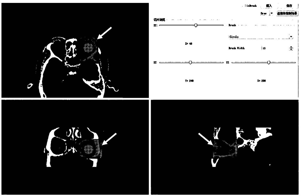
[0067] The digital model of the prosthetic eye seat is obtained through CT scanning of the medical digital imaging and communication (DICOM) data of the orbital bony structure and soft tissue in the patient's eye and healthy eye, and the medical digital imaging of the orbital bony structure and soft tissue structure in the orbit Segmentation, three-dimensional modeling and analysis of data and communication (DICOM) to construct and obtain individualized digital models.
[0068] The specific implementation takes the 3DMed medical image processing and analysis system as the development platform, develops the image segmentation algorithm as the core, and constructs an interactive image segmentation method.
[0069] The specific construction method of the digital model of the prosthetic eye seat is as follows:
[0070] 1) ...
Embodiment 2
[0101] Embodiment 2: animal experiments
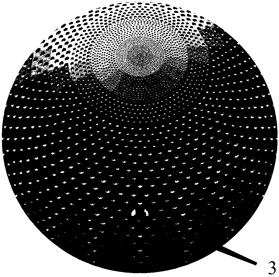
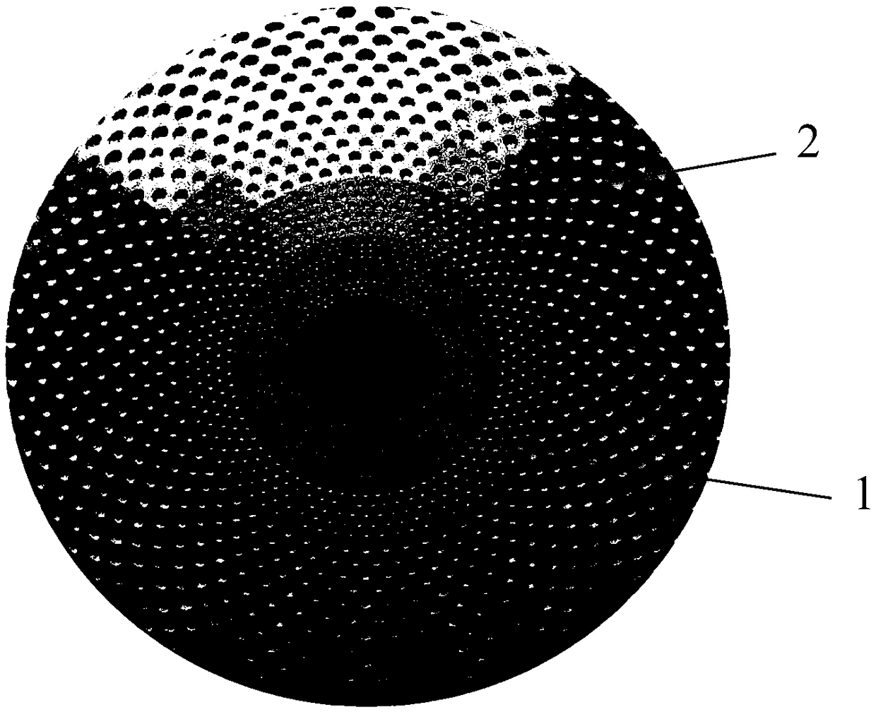
[0102] The 3D printed hydroxyapatite prosthetic eye socket prepared in Example 1 was used as the experimental group, and the hydroxyapatite eye socket scaffold made of pore-forming agent was used as the control group to evaluate the vascularization efficiency and eye socket mobility of the eye socket material . The result is as Figure 6 Shown: The 3D printed hydroxyapatite prosthetic eye seat has a higher density of neovascularization than that of the control group (A is the control group, B is the experimental group, and the arrow shows the neovascularization). Figure 7 Shown is the range of movement of the eye socket in the upward, downward, left and right directions after implantation of the artificial eye socket, which shows that the artificial eye socket has good mobility.
PUM
 Abstract
Description
Claims
Application Information