Optical ultrasound imaging device for diagnosing breast cancer

An imaging device, ultrasonic technology, applied in the direction of diagnosis, diagnostic recording/measurement, medical science, etc., can solve the problems that cannot be applied to the uneven surface of the human body, and the number of detection equipment probes is small, and achieve simple and portable structure, high sensitivity, and device simple effect

Inactive Publication Date: 2019-01-04
NANCHANG HANGKONG UNIVERSITY
View PDF5 Cites 6 Cited by
  • Summary
  • Abstract
  • Description
  • Claims
  • Application Information

AI Technical Summary

Problems solved by technology

[0004] Second: Most of the current detection equipment is general-purpose equipment, which cannot be applied to some uneven surfaces of the human body;
[0005] Third: The number of ultrasonic probes directly determines the number of echoes used for post-imaging, and the current detection equipment has a small number of probes

Method used


Image

  • Optical ultrasound imaging device for diagnosing breast cancer
  • Optical ultrasound imaging device for diagnosing breast cancer
  • Optical ultrasound imaging device for diagnosing breast cancer

Examples


Embodiment Construction

[0027] In order to make the object, technical solution and advantages of the present invention clearer, the present invention will be further described in detail below in conjunction with the accompanying drawings and specific embodiments. It should be understood that the specific embodiments described here are only used to explain the present invention, and are not intended to limit the present invention.

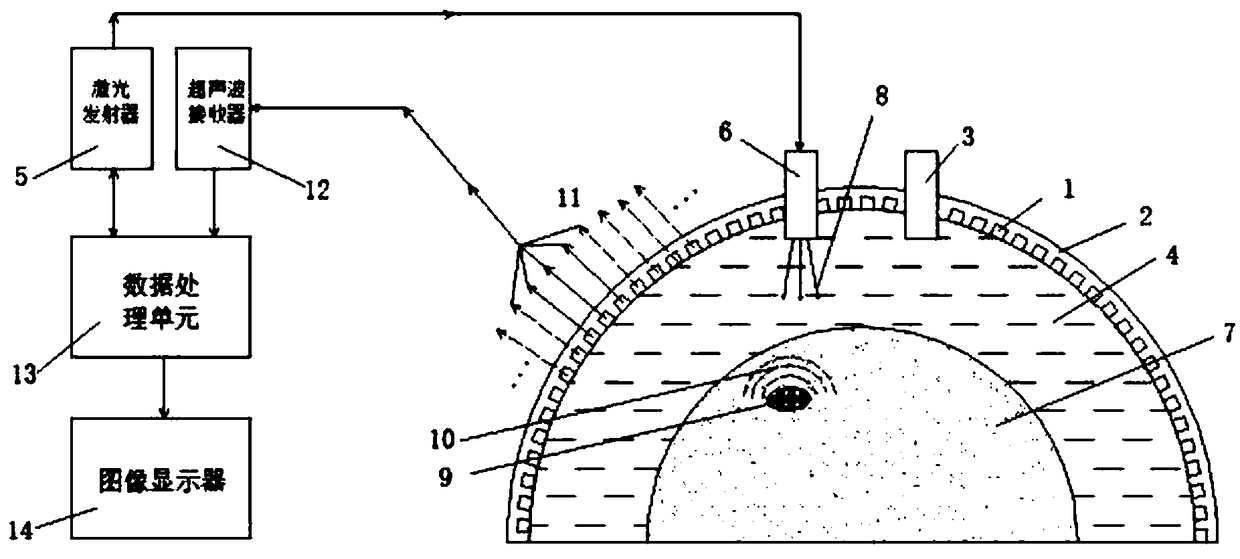
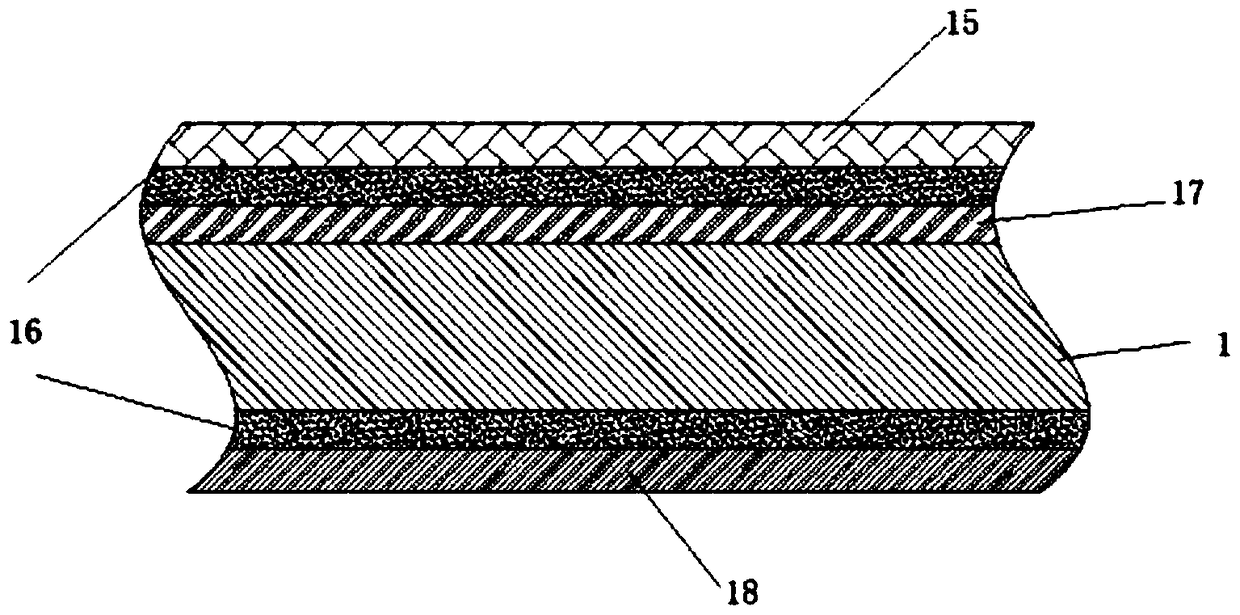
[0028] see figure 1 , the specific implementation mode adopts the following technical solutions: a bowl-shaped flexible photoacoustic imaging device, the composite chip 1 is installed on the bowl-shaped flexible structure 2 according to the rules to form an array structure. The upper end of the flexible structure is equipped with a couplant injection port 3, and the bowl-shaped probe is placed on the patient during work, and the edge is closely attached to the surface of the human body. The coupling agent 4 is injected from the coupling agent injection port 3, and the edg...


PUM


Abstract

The invention discloses an optical ultrasonic imaging device for diagnosing breast cancer, which comprises a bowl-shaped flexible ultrasonic probe, a laser probe, a pulsed laser emitter, an ultrasonicreceiver, a data processing unit and an image display. The device comprises a bowl-shaped flexible ultrasonic probe, a laser probe, a pulsed laser emitter, an ultrasonic receiver, a data processing unit and an image display. The ultrasonic transducer adopts bowl-shaped shell and flexible structure, adopts multi-array array composite wafer structure, utilizes multi-array array composite wafer, andcombines the principle of phased array technology to obtain the signal sources with different initial phases. An ultrasonic transducer is connected to an ultrasonic receiver. A laser probe connectedwith a pulse laser emitter emits a laser beam with a certain frequency to that inside of the tissue, molecules or substances that absorb light at a particular wavelength are called chromophores. The light energy absorbed by the chromophore is converted into heat energy by molecular vibration and thermoelastic expansion to generate ultrasonic wave, which is detected by an ultrasonic sensor in the device, and the waveform signal is synthesized into an image through an ultrasonic receiver and a data processing unit and displayed on an image display.

Description

technical field [0001] The invention relates to the field of medical breast cancer lesion diagnosis and adjuvant therapy, in particular to an optical ultrasonic imaging device for breast cancer diagnosis that can detect superficial (in vivo) lesions. Background technique [0002] When the pulsed laser irradiates the biological tissue, the absorber located in the tissue absorbs the pulsed light energy, generates instantaneous temperature rise and expands, and generates ultrasonic waves. At this time, the ultrasonic detector located on the surface of the tissue body can receive these external ultrasonic waves, and reconstruct the image of the light energy absorption distribution in the tissue according to the detected photoacoustic signal. After the laser beam is incident on the tissue surface, the penetration depth of the beam varies according to the wavelength of the laser. In this process, the light beam is subjected to multiple scattering and absorption, and molecules or ...

Claims


Application Information

Patent Timeline
Patent Type & Authority Applications(China)
IPC IPC(8): A61B5/00
CPCA61B5/0095
Inventor 常俊杰李媛媛王焜
Owner NANCHANG HANGKONG UNIVERSITY
Why Eureka
Try Eureka