
Human cell whole form immunofluorescence staining method and kit

An immunofluorescence staining and human cell technology, applied in the field of human cell full-form immunofluorescence staining methods and kits, can solve the problem of inability to distinguish nucleolus and nucleoplasm, low sensitivity of nucleolar color development, and inability to clearly visualize nucleoli. shape and structure

Active Publication Date: 2021-03-30
青岛言鼎生物医疗科技有限公司
View PDF5 Cites 0 Cited by
  • Summary
  • Abstract
  • Description
  • Claims
  • Application Information

AI Technical Summary

Problems solved by technology

Its main technical disadvantages are that it cannot clearly visualize the shape and structure of nucleoli, cannot clearly mark nucleoli, and cannot distinguish nucleoli from nucleoplasm; the chemical staining and histological examination methods used in exfoliated cells (including cervical exfoliated cells) are basically Similarly, it can display the basic structure of the cell, and can display the nucleus, but cannot clearly display the morphological structure of the nucleolus, cannot calibrate the nucleolus, and cannot distinguish between the nucleolus and the nucleoplasm
[0006] Bone marrow cytology uses chemical staining, which can visualize the overall structure of cells and display nuclei and nucleoli, but bone marrow cytology uses general chemical staining, which is less sensitive to nucleolus color development, and cannot distinguish nucleoli from nucleoli. Plasma, which brings certain uncertainty to the cytological diagnosis of blood diseases
[0007] The current chemical staining for CTC cytology detection has poor reproducibility and low sensitivity, and basically cannot clearly display the morphological structure of the nucleolus; the mainstream immunofluorescent staining method focuses on displaying the antigens on the cell surface, but cannot display the nucleolus The morphology and structure of the cell can be judged by the antigen or DAPI stained nucleus to determine whether it is a CTC cell. False positives or false negatives may occur due to changes in antigens or illegal editing of antigens
Inability to examine and identify CTCs from cytopathological features

Method used


Image

  • Human cell whole form immunofluorescence staining method and kit

Examples


Embodiment 1

[0084] Example 1: Cell characteristic direct immunofluorescence cell staining process

[0085] Main reagents:

[0086] ① Fluorescent dye (Alexa 647) labeled nucleolin (Nucleolin), orange red;

[0087] ② Fluorescent dye (Alexa 594) Marked nucleolar fibrin (fibrilarin), red;

[0088] ③Nuclear fluorescent dye: DAPI, blue-purple;

[0089] ④ fluorescent dye (Alexa 488) labeled anti-CK7, CK18, CK19 monoclonal antibody, green;

[0090] Pay attention to the wavelength and color matching of each fluorescent dye.

[0091] Cell fixatives: methanol, acetone, paraformaldehyde.

[0092] The cell blocking solution includes: pH 7.2-7.4 PBS (containing 0.5-10.0% BSA or calf serum).

[0093] The washing solution includes: pH7.2-7.4PBS (containing Tween-20).

[0094] The diluent includes: pH 7.2-7.4 PBS (containing 0.1-1.5% BSA or calf serum), adding appropriate concentration of fluorescent labeled antibody or non-labeled antibody to form antibody binding reaction solution.

[0095]...

Embodiment 2

[0107] Example 2: Cell characteristic indirect immunofluorescence cell staining process

[0108] Main reagents:

[0109] ① Nucleolin antibody.

[0110] ② nucleolar fibrin (fibrillarin) antibody.

[0111] ③Nuclear fluorescent dye: DAPI, blue-purple.

[0112] ④ fluorescent dye (Alexa 488) labeled anti-CK7, CK18, CK19 monoclonal antibody, green.

[0113] ⑤Apoptosis susceptibility protein antibody—goat anti-rabbit (Alexa 488) Fluorescently labeled antibodies.

[0114] ⑥ Goat anti-rabbit IgG (Alexa 594) labeled fluorescent secondary antibody.

[0115] Pay attention to the wavelength and color matching of each fluorescent dye.

[0116] Cell fixatives: methanol, acetone, paraformaldehyde.

[0117] The cell blocking solution includes: pH 7.2-7.4 PBS (containing 0.5-10.0% BSA or calf serum).

[0118] The washing solution includes: pH7.2-7.4PBS (containing Tween-20).

[0119] The diluent includes: pH 7.2-7.4 PBS (containing 0.1-1.5% BSA or calf serum), adding appropriate c...

Embodiment 3

[0133] Example 3: Human blood cell staining

[0134] Main reagents:

[0135] ① Fluorescent dye (Alexa 647) labeled nucleolin (Nucleolin), orange red.

[0136] ② Fluorescent dye (Alexa 594) labeled fibrillin, red.

[0137] ③Nuclear fluorescent dye: DAPI, blue-purple.

[0138] ④ fluorescent dye (Alexa 488) Monoclonal antibody labeled CD45, (leukocytes) in green.

[0139] Pay attention to the wavelength and color matching of each fluorescent dye.

[0140] Cell fixatives: methanol, acetone, paraformaldehyde.

[0141] The cell blocking solution includes: pH 7.2-7.4 PBS (containing 0.5-10.0% BSA or calf serum).

[0142] The washing solution includes: pH7.2-7.4PBS (containing Tween-20).

[0143] The diluent includes: pH 7.2-7.4 PBS (containing 0.1-1.5% BSA or calf serum), adding appropriate concentration of fluorescent labeled antibody or non-labeled antibody to form antibody binding reaction solution.

[0144] Cell membrane and nuclear membrane permeabilization solution...


PUM


Abstract

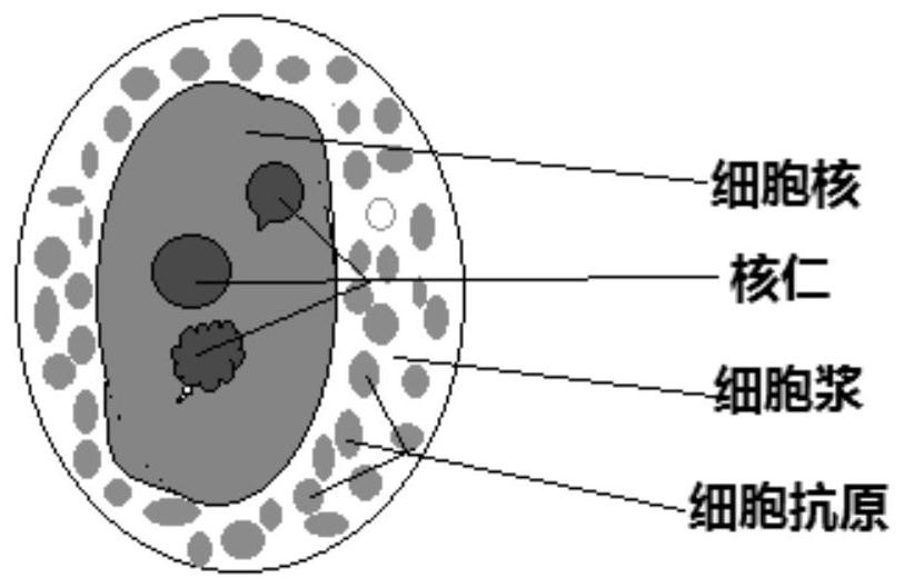
The invention discloses a human cell holomorphic immunofluorescent staining method and a kit. Human cells are subjected to holomorphic staining by direct immunofluorescence assay or indirect immunofluorescence assay by using antibodies of cell nuclear antigens, cell nucleolus antigens and nucleoplasm antigens and antibodies of related antigens in cytomembrane and cytoplasm. The cell nucleus, particularly nucleolus capable of calibrating the nucleus can be clearly and completely displayed, and the quantity, form, size and distribution of the nucleolus are clearly visible. In addition to characteristic display of the antigens of the cells by immunofluorescent staining, the human cells can be subjected to holomorphic observation and analysis.

Description

technical field [0001] The present invention generally relates to the field of medical detection and analysis, in particular to a method and kit for immunofluorescent staining of human cells in whole form. Background technique [0002] Human cells are generally divided into three parts: cell membrane, cytoplasm (cytoplasm) and nucleus, but in the pathological examination of malignant cells, special attention is paid to the pathological and morphological changes of the nucleus of malignant cells and the nucleolus in the nucleus. The nucleolus in the nucleus is a vital organelle in the nucleus and is the hub of all cell activities. The change of the nucleus is particularly critical, and the change of the nucleolus is the pathological basis and core evidence for identifying malignant cells. [0003] Under the electron microscope, the structure of the nucleolus in the nucleus is divided into: (1) fiber center: the fiber center is the innermost structure of the nucleolus, accoun...

Claims


Application Information

Patent Timeline
Patent Type & Authority Patents(China)
IPC IPC(8): G01N1/30G01N33/58G01N33/533
CPCG01N1/30G01N33/533G01N33/582G01N2001/302
Inventor 肖乐义刘元柱米明仁杨勤英李娟
Owner 青岛言鼎生物医疗科技有限公司
Learn More