Method and application for systematically freezing and storing human umbilical cord tissues according to structure levels

A human umbilical cord and cryopreservation technology, applied in the field of life sciences, can solve the problems of sample resource waste, high preparation cost, and long cycle, and achieve the effects of maintaining integrity, increasing cell yield, and improving cryopreservation and resuscitation activity

Active Publication Date: 2019-03-01
YINFENG BIOLOGICAL GRP
View PDF9 Cites 3 Cited by
  • Summary
  • Abstract
  • Description
  • Claims
  • Application Information

AI Technical Summary

Problems solved by technology

This method has a long period of time and high preparation costs, and other types of tissues in the umbilical cord and the remaining Wharton jelly tissues will be discarded as waste, resulting in a waste of sample resources

Method used


Image

  • Method and application for systematically freezing and storing human umbilical cord tissues according to structure levels
  • Method and application for systematically freezing and storing human umbilical cord tissues according to structure levels
  • Method and application for systematically freezing and storing human umbilical cord tissues according to structure levels

Examples


Embodiment 1

[0050] The cryopreservation of embodiment 1 people's umbilical cord tissue

[0051] Proceed as follows:

[0052] (1) Wash the umbilical cord 3 times with normal saline, cut off both ends of the umbilical cord with tissue scissors, rinse twice with normal saline, tear off the surface amniotic tissue (try to keep the amniotic tissue intact), and remove the amniotic tissue, each less than 10cm 2 ;Rinse the amnion tissue with normal saline, fold the amnion or place it flat in a cryopreservation bag, add the balance solution pre-cooled to 4°C, balance for 8 minutes, centrifuge at 4°C, 1300rpm for 5 minutes, remove the balance solution, and then add the balance solution pre-cooled to 4°C ℃ vitrification solution Ⅰ, transferred to liquid nitrogen for cryopreservation;

[0053] The freezing solution includes 1) base solution: α-MEM medium containing 20wt% human serum albumin; 2) balance solution: adding 20wt% ethylene glycol to the base solution; 3) vitrification solution: basic 55...

Embodiment 2

[0080] Example 2: Recovery after umbilical cord cryopreservation

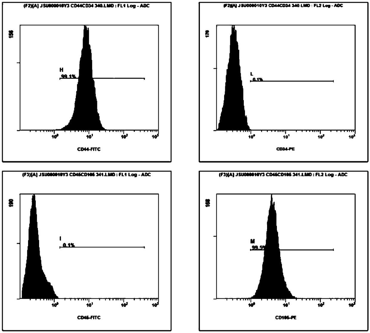
[0081] After cryopreservation for 3 months according to the method of Example 1, resuscitation was carried out, and mesenchymal stem cells were induced and isolated. The steps were as follows:

[0082] (1) Recovery of frozen umbilical cord amniotic tissue: Take out the frozen umbilical cord amniotic tissue from liquid nitrogen, put the cryopreservation bag in a water bath at 37°C to quickly dissolve for 80 seconds (about 80% dissolved), and then quickly transfer Put it in a biological safety cabinet, open the cryopreservation bag, gently remove the amniotic membrane with tweezers, put it into a centrifuge tube (50ml), add resuscitation solution Ⅰ (equilibrium at room temperature in advance), mix well, centrifuge at low temperature, remove the supernatant; then add Resuscitation solution II (balanced at room temperature in advance), mix well, centrifuge at low temperature, remove supernatant, and set aside;

[...


PUM


Abstract

The invention discloses a method for systematically freezing and storing human umbilical cord tissues according to structure levels. The method comprises the following steps of (1) separation and freeze storage of umbilical cord amniotic membrane tissues; (2) separation and freeze storage of umbilical cord blood vessel tissues; (3) separation and freeze storage of umbilical cord Wharton's jelly tissues. The invention also provides an umbilical cord restoration method. The freeze storage method provided by the invention has the advantages that the umbilical cord amniotic membrane, blood vesselsand Wharton's jelly tissues are sequentially peeled according to the structure levels; then through treatment, the materials are put into liquid nitrogen for freeze storage; three tissues of the umbilical cord are respectively stored; different freeze storage and restoration methods are used according to the specificity of each tissue; the freeze storage restoration activity and cell yield are favorably improved. The invention provides a set of methods for systematically storing complete umbilical cord tissues of various types; important biological resources are provided for the study of thestem cells from the umbilical cord and can be used for building tissue or cell resource sample databases derived from the umbilical cord. The method of the invention provides a solid foundation and basis for completing the human genetic resource databases from the umbilical cord.

Description

technical field [0001] The invention relates to a method and application for systematically freezing and storing human umbilical cord tissue according to structure levels, and belongs to the technical field of life sciences. Background technique [0002] The umbilical cord is a tubular structure that connects the fetus and the placenta in mammals. The outer layer is amniotic membrane, and the inner layer is composed of two arteries and a vein. In the gap, loose and jelly-like mesenchyme can be seen, filled with Wharton's jelly tissue , can isolate and prepare a variety of stem cells, and is a rich tissue resource bank. [0003] Human amniotic epithelial cells (hAEC) can be isolated from amniotic membrane tissue, which has the characteristics of expressing multiple embryonic stem cell markers and has a relatively comprehensive multi-lineage differentiation potential. In addition to expressing embryonic stem cell surface antigens: SSSEA-3 and SSSEA-4, tumor resistance genes T...

Claims


Application Information

Patent Timeline
Patent Type & Authority Applications(China)
IPC IPC(8): A01N1/02C12N5/0775
CPCA01N1/0221C12N5/0662
Inventor 崔光晶徐峰波王圣川宋现收王肇光生德伟
Owner YINFENG BIOLOGICAL GRP
Why Eureka
Try Eureka