A kind of exosome with diagnosis and treatment function and preparation method thereof
A technology of exosomes and functions, applied in the field of nanomedicine, can solve the problems of insufficient timeliness and accuracy in tumor diagnosis and condition evaluation, and achieve the effects of improving solubility, short preparation time, and easy operation
- Summary
- Abstract
- Description
- Claims
- Application Information
AI Technical Summary
Problems solved by technology
Method used
Image
Examples
Embodiment 1
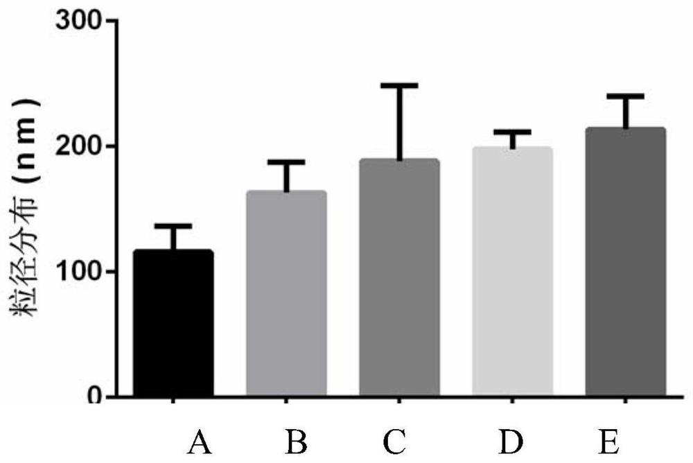
[0043] Add 80 μg of exosomes to 10 mM PBS solution, mix well, add to a 96-well plate, put it into an electroporation instrument, and operate according to the following parameters: voltage 250 V, pulse width 100 μs, interval 1000 μs, discharge times 6 times. After electroporation, put the suspension in a cell culture incubator and incubate for 30 minutes, then ultracentrifuge twice at 100,000g for 70 minutes each time, remove the supernatant, and resuspend the pellet with 200 μL PBS to obtain exosomes after electroporation. Its particle size is ~150nm, such as image 3 shown. Zeta potential is ~-8mV, such as Figure 4 shown. Store at 4°C in the dark for subsequent testing and experiments.
Embodiment 2
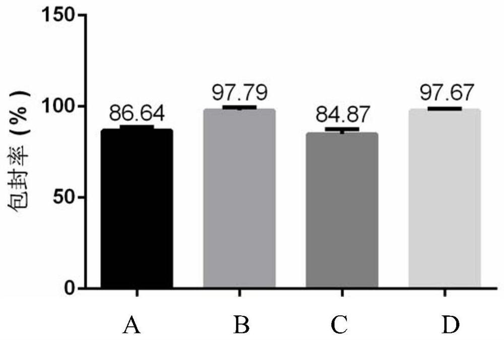
[0045] Add 80 μg of exosomes and 80 μL of curcumin solution (concentration: 2 mg / mL) into 10 mM PBS solution, mix well, add to a 96-well plate, put it into an electroporation apparatus, and operate according to the following parameters: voltage 250 V, pulse width 100 μs , the interval is 1000μs, and the number of discharges is 6 times. After electroporation, put the suspension in a cell culture incubator and incubate for 30 minutes, then ultracentrifuge twice at 100,000 g for 70 minutes each time, remove the supernatant, and resuspend the pellet with 200 μL PBS to obtain curcumin-loaded exosomes. Its encapsulation efficiency is ~86.64%, such as figure 2 Shown; its particle size is ~ 170nm, such as image 3 Shown; Zeta potential is ~-17mV, such as Figure 4 shown; the prepared exosomes loaded with curcumin have drug sustained release properties, such as Image 6 shown. Store at 4°C in the dark for subsequent testing and experiments.
Embodiment 3
[0047] Add 80 μg of exosomes and 40 μL of indocyanine green solution (concentration: 2 mg / mL) into 10 mM PBS solution, mix well, add to a 96-well plate, put it into an electroporation apparatus, and operate according to the following parameters: voltage 250 V, pulse The width is 100μs, the interval is 1000μs, and the number of discharges is 6 times. After electroporation, place the suspension in a cell culture incubator and incubate for 30 minutes, then ultracentrifuge twice at 100,000 g for 70 minutes each time, remove the supernatant, and resuspend the pellet with 200 μL PBS to obtain indocyanine green-loaded exocytosis. body, its encapsulation efficiency is ~97.79%, such as figure 2 Shown; the particle size is ~180nm, such as image 3 Shown; Zeta potential is ~-6mV, such as Figure 4 shown. The prepared exosomes loaded with indocyanine green have fluorescence imaging, photothermal effect and drug sustained release properties, such as figure 1 , 5, 6. Store at 4°C in t...
PUM
| Property | Measurement | Unit |
|---|---|---|
| diameter | aaaaa | aaaaa |
| particle diameter | aaaaa | aaaaa |
| particle diameter | aaaaa | aaaaa |
Abstract
Description
Claims
Application Information
 - R&D
- Intellectual Property
- Life Sciences
- Materials
- Tech Scout
- Unparalleled Data Quality
- Higher Quality Content
- 60% Fewer Hallucinations
Browse by: Latest US Patents, China's latest patents, Technical Efficacy Thesaurus, Application Domain, Technology Topic, Popular Technical Reports.
© 2025 PatSnap. All rights reserved.Legal|Privacy policy|Modern Slavery Act Transparency Statement|Sitemap|About US| Contact US: help@patsnap.com


