
Method for inducing dental pulp stem cells (DPSCs) to be differentiated into cardiomyocyte-like cells

A technique for dental pulp stem cells and cardiomyocyte-like cells, applied in the field of stem cells, can solve problems such as no DPSCs, and achieve the effects of avoiding immune rejection, easy in vitro expansion, and convenient material sampling.

Inactive Publication Date: 2019-04-05
GUANGZHOU SALIAI STEMCELL SCI & TECH CO LTD
View PDF3 Cites 0 Cited by
  • Summary
  • Abstract
  • Description
  • Claims
  • Application Information

AI Technical Summary

Problems solved by technology

At present, there are few reports on the induction of DPSCs differentiation into cardiomyocytes, and there is no report on the effective induction of DPSCs differentiation into cardiomyocytes

Method used


Image

  • Method for inducing dental pulp stem cells (DPSCs) to be differentiated into cardiomyocyte-like cells
  • Method for inducing dental pulp stem cells (DPSCs) to be differentiated into cardiomyocyte-like cells
  • Method for inducing dental pulp stem cells (DPSCs) to be differentiated into cardiomyocyte-like cells

Examples


Embodiment 1

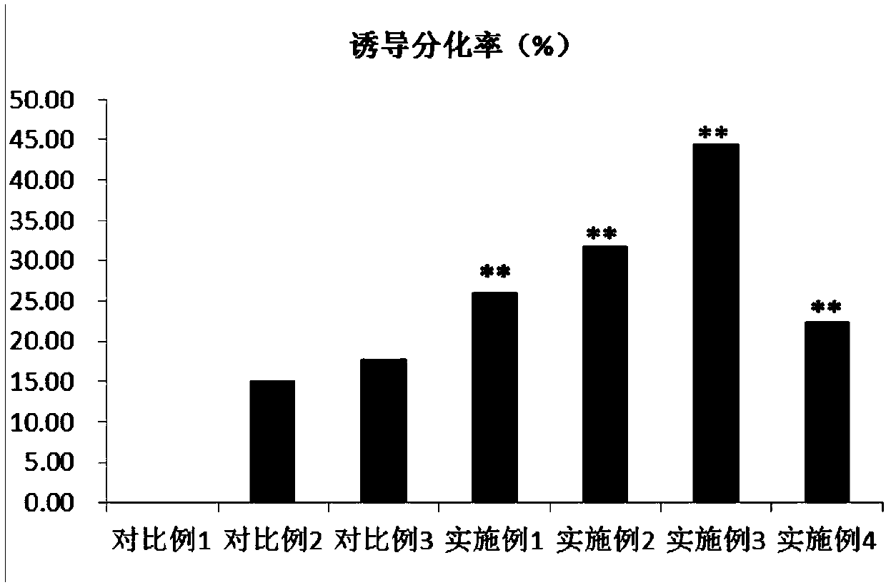
[0041] Select the 3rd generation DPSCs, press 1×10 4 Cell / mL density was inoculated in a six-well plate with polylysine-treated sterile coverslips, and 2ml of DMEM / F12 medium containing 10% FBS and 10ng / ml EGF was added to each well, and placed at 37°C , 5%CO 2 After 24 hours of cultivation, replace the DMEM / F12 medium containing 10% FBS, 5 μmol / L 5-aza, and 10 μmol / L PFT-α; after 24 hours of induction culture, replace the DMEM / F12 medium containing 15% FBS The F12 medium was maintained for 4 weeks, and new medium was replaced every 2-3 days. After 4 weeks of culture, the cell slides were taken out, and the immunohistochemistry of desmin, troponin I and troponin T was performed according to the instructions of the immunohistochemical staining kit. Staining, DAB color development.

Embodiment 2

[0043] Select the 3rd generation DPSCs, press 1×10 4 Cell / mL density was inoculated in a six-well plate with polylysine-treated sterile coverslips, and 2ml of DMEM / F12 medium containing 10% FBS and 10ng / ml EGF was added to each well, and placed at 37°C , 5%CO 2 After 24 hours of cultivation, replace the DMEM / F12 medium containing 10% FBS, 10 μmol / L 5-aza, and 15 μmol / L PFT-α; after 24 hours of induction culture, replace the DMEM / F12 medium containing 15% FBS The F12 medium was maintained for 4 weeks, and new medium was replaced every 2-3 days. After 4 weeks of culture, the cell slides were taken out, and the immunohistochemistry of desmin, troponin I and troponin T was performed according to the instructions of the immunohistochemical staining kit. Staining, DAB color development.

Embodiment 3

[0045] Select the 3rd generation DPSCs, press 1×10 4 Cell / mL density was inoculated in a six-well plate with polylysine-treated sterile coverslips, and 2ml of DMEM / F12 medium containing 10% FBS and 10ng / ml EGF was added to each well, and placed at 37°C , 5%CO 2 After 24 hours of culture, replace the DMEM / F12 medium containing 10% FBS, 10 μmol / L 5-aza, and 20 μmol / L PFT-α; after 24 hours of induction culture, replace the DMEM / F12 medium containing 15% FBS The F12 medium was maintained for 4 weeks, and new medium was replaced every 2-3 days. After 4 weeks of culture, the cell slides were taken out, and the immunohistochemistry of desmin, troponin I and troponin T was performed according to the instructions of the immunohistochemical staining kit. Staining, DAB color development.


PUM


Abstract

The invention belongs to the field of stem cells and discloses a composition and a method for inducing dental pulp stem cells (DPSCs) to be differentiated into cardiomyocyte-like cells. The composition for inducing the dental pulp stem cells to be differentiated into the cardiomyocyte-like cells, provided by the invention, is prepared from 5-azacytidine and PFT-a. The method for inducing the dental pulp stem cells to be differentiated into the cardiomyocyte-like cells, provided by the invention, comprises the following steps: after pre-culturing the dental pulp stem cells, replacing with a DMEM (Dulbecco's Modified Eagle Medium) / F12 culture medium containing FBS (Fetal Bovine Serum) and the composition for inducing the dental pulp stem cells to be differentiated into the cardiomyocyte-likecells and carrying out inducing culture; then replacing with a DMEM / F12 culture medium containing the FBS and maintaining the culture. The autologous dental pulp stem cells are convenient to obtain,have an abundant source, are easy to amplify in vitro and can be used for autoplastic transplantation, so that the immune rejection response is avoided. An experiment shows that two inducing factors including the 5-azacytidine and the PFT-a are combined and applied to the dental pulp stem cells and the DPSCs can be effectively induced to be differentiated into the cardiomyocyte-like cells.

Description

technical field [0001] The invention belongs to the field of stem cells, and in particular relates to a method for inducing differentiation of dental pulp stem cells into cardiomyocyte-like cells. Background technique [0002] Myocardial infarction is the myocardial damage caused by the imbalance between coronary blood flow and myocardial demand caused by changes in coronary circulation. It is a serious clinical ischemic heart disease. Cardiomyocytes after necrosis are gradually replaced by scar tissue. Due to the lack of elasticity of scar tissue, it is difficult to meet the requirements of cardiac contraction and relaxation. In order to compensate for the loss of cardiomyocyte function, the heart gradually undergoes degenerative left ventricular remodeling. decline, eventually leading to congestive heart failure and even sudden death. Although clinical drugs and interventional therapy can improve symptoms, they cannot restore damaged cardiomyocytes. Although heart transp...

Claims


Application Information

Patent Timeline
IPC IPC(8): C12N5/077
CPCC12N5/0657C12N2500/40C12N2501/48C12N2506/1361
Inventor 陈海佳葛啸虎戚康艺王小燕
Owner GUANGZHOU SALIAI STEMCELL SCI & TECH CO LTD
Learn More