Exosome surface protein detection method

A surface protein and detection method technology, applied in the direction of measuring device, color/spectral characteristic measurement, instrument, etc., can solve the problems of high cost, complex detection operation of exosome surface protein, low sensitivity, etc., to improve reliability, fast The effect of highly sensitive exosome surface protein detection and simplified detection steps

Inactive Publication Date: 2019-04-16
UNIV OF SHANGHAI FOR SCI & TECH
View PDF0 Cites 9 Cited by
  • Summary
  • Abstract
  • Description
  • Claims
  • Application Information

AI Technical Summary

Problems solved by technology

[0005] The purpose of the present invention is to provide a method for detecting exosome surface proteins, which can detect exosome surface proteins through colorimetry and SERS spectrum technology dual-signal, on the one hand, through colorimetry, can realize the rapid characterization of exosome surface proteins Detection, on the other hand, through the confocal Raman microscopy system to analyze the SERS signal technology in the product, the precise quantitative analysis of exosome surface proteins solves the problem of complex detection operations, low sensitivity, and low sensitivity of exosome surface proteins in the prior art. high cost

Method used


Image

  • Exosome surface protein detection method

Examples


preparation example Construction

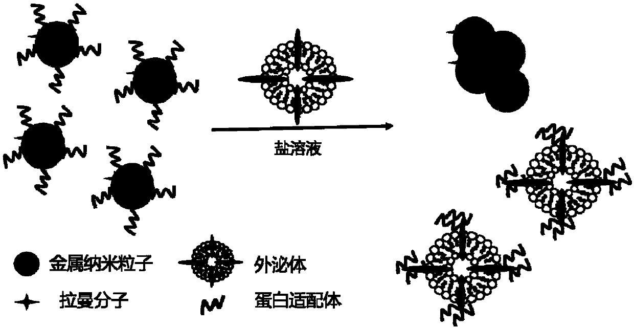
[0038] The preparation method of described reporter probe, comprises the steps:

[0039] The first step, adopting sodium citrate reduction method to prepare spherical gold nanoparticles;

[0040] In the second step, Raman molecules are connected on the surface of the spherical gold nanoparticles prepared in the first step.

Embodiment 1

[0042] Using DTNB as the Raman molecule, human breast cancer cell SKBR3 and human cervical cancer cell HeLa as the source of exosomes, the method of the present invention was used to detect the HER2 protein on the surface of the exosomes.

[0043] Step 1, preparing spherical gold nanoparticles:

[0044] Add 200uL of 10% HAuCl in 200mL deionized water 4 solution, stirring vigorously and heating to boiling. Then add 8 mL of 1% sodium citrate aqueous solution, and continue heating and stirring for 15 min. Stop heating, homogeneously stir the solution and cool it down to room temperature to obtain a wine-red spherical gold nanoparticle solution;

[0045] Step 2, connect DTNB to the surface of the spherical gold nanoparticles obtained in step 1 to obtain the reporter probe:

[0046] Take 200 μL of the spherical gold nanoparticle solution in step 1, then add 4 μL of 10 mM DTNB solution, and continue to stir the mixture for 2 hours, then centrifuge and wash twice with distilled wate...

Embodiment 2

[0057] Using DTNB as the Raman molecule and the source of human breast cancer cell SKBR3 exosomes, the quantitative detection experiment of exosome surface proteins was carried out by using the method of the present invention.

[0058] Prepare the reporter probe according to steps 1 and 2 in Example 1, and extract SKBR3 exosomes according to the method described in Step 3 in Example 1. In the detection experiment, add different volumes of SKBR3 exosome solutions, respectively 10 and 40 , 60, 80 μL, the corresponding SKBR3 exosome concentrations are 0.09nM, 0.36nM, 0.54nM, 0.72nM, the blank control group uses PBS solution, the rest of the steps refer to Example 1, and finally observe its color or detect its SERS signal.

[0059] The SERS spectra of the reporter probes obtained in this example reacted with different concentrations of SKBR3 exosomes. With the increase of the concentration of SKBR3 exosomes added, the SERS signal of the reporter probes also increased continuously. ...


PUM

PropertyMeasurementUnit
sizeaaaaaaaaaa
Sensitivityaaaaaaaaaa

Abstract

The invention discloses an exosome surface protein detection method, which comprises the steps of first, extracting exosomes from a tumor cell culture medium by centrifugation; second, using a reportprobe to react with an exosome to be detected, wherein the report probe is a spherical gold nanoparticle, and the surface is connected with a protein adapter and a Raman molecular; and third, observing the color of the report probe or detecting an SERS signal of the report probe. According to the invention, a colorimetric method and an SERS detection technology are simultaneously applied to the exosome surface protein detection system, firstly the sample is quickly analyzed by adopting the colorimetric method, and then the SERS technology is utilized to achieve accurate quantitative analysis for the exosome surface protein, so that fast and high-sensitivity detection for the exosome surface protein is realized by combining the two methods.

Description

technical field [0001] The invention belongs to the field of exosome surface protein detection, and in particular relates to a method for detecting exosome surface protein. Background technique [0002] Exosomes are vesicles secreted by cells with a size of about 30-120nm. The outer layer is a lipid membrane. Specific proteins are expressed on the surface of the membrane, and the interior contains biomolecules such as proteins, DNA, and RNA. Since exosomes are secreted by host cells, the surface proteins and contents of exosomes can reflect the physiological state and information of secreting cells. Studies have found that the exosomes of tumor cells are closely related to the development of tumors, and many tumor cell surface markers are also expressed on the surface of exosomes. Therefore, the detection of exosome surface proteins can replace the tissue biopsy of tumor cells to a certain extent. This makes exosomes an important tumor marker and therapeutic target. The de...

Claims


Application Information

Patent Timeline
Patent Type & Authority Applications(China)
IPC IPC(8): G01N21/65G01N21/25
CPCG01N21/25G01N21/658
Inventor 陈辉马佩李钧颖张学典
Owner UNIV OF SHANGHAI FOR SCI & TECH
Why Eureka
Try Eureka