A lung lobe segmentation method, device, computer equipment and storage medium

A lung lobe and image segmentation technology, applied in the field of devices, lung lobe segmentation methods, computer equipment and storage media, can solve the problems of the precise influence of automatic lung lobe segmentation results and cannot meet clinical needs, and achieve the effect of improving accuracy

Active Publication Date: 2021-12-28
SHANGHAI UNITED IMAGING HEALTHCARE
View PDF19 Cites 0 Cited by
  • Summary
  • Abstract
  • Description
  • Claims
  • Application Information

AI Technical Summary

Problems solved by technology

However, the quality of lung CT image data and the accuracy of the automatic lung lobe segmentation algorithm will have a certain impact on the accuracy of the automatic lung lobe segmentation results, so that the results of the automatic lung lobe segmentation cannot meet the clinical needs, and further optimization of the automatic lung lobe segmentation results is required

Method used


Image

  • A lung lobe segmentation method, device, computer equipment and storage medium
  • A lung lobe segmentation method, device, computer equipment and storage medium
  • A lung lobe segmentation method, device, computer equipment and storage medium

Examples


Embodiment 1

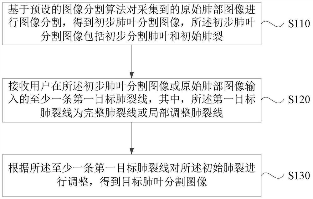
[0068] figure 1 It is a flow chart of the lung lobe segmentation method provided in Embodiment 1 of the present invention. This embodiment is applicable to the case of processing lung medical images, and the method can be executed by a lung lobe segmentation device.

[0069] Such as figure 1 As shown, the lung lobe segmentation method specifically includes the following steps:

[0070] S110. Perform image segmentation on the acquired original lung image based on a preset image segmentation algorithm to obtain a preliminary lung lobe segmentation image, and the preliminary lung lobe segmentation image includes a preliminary segmented lung lobe and an initial lung fissure.

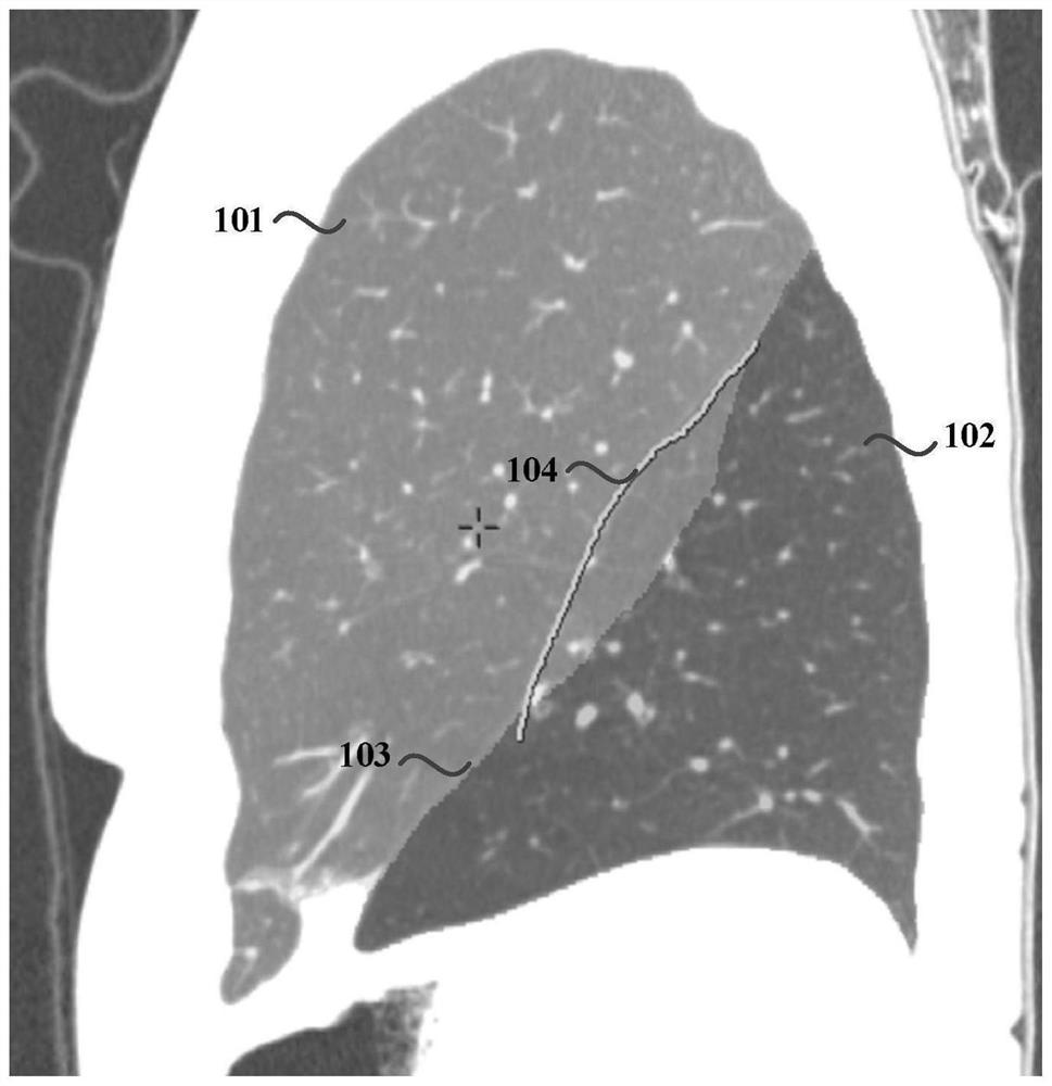
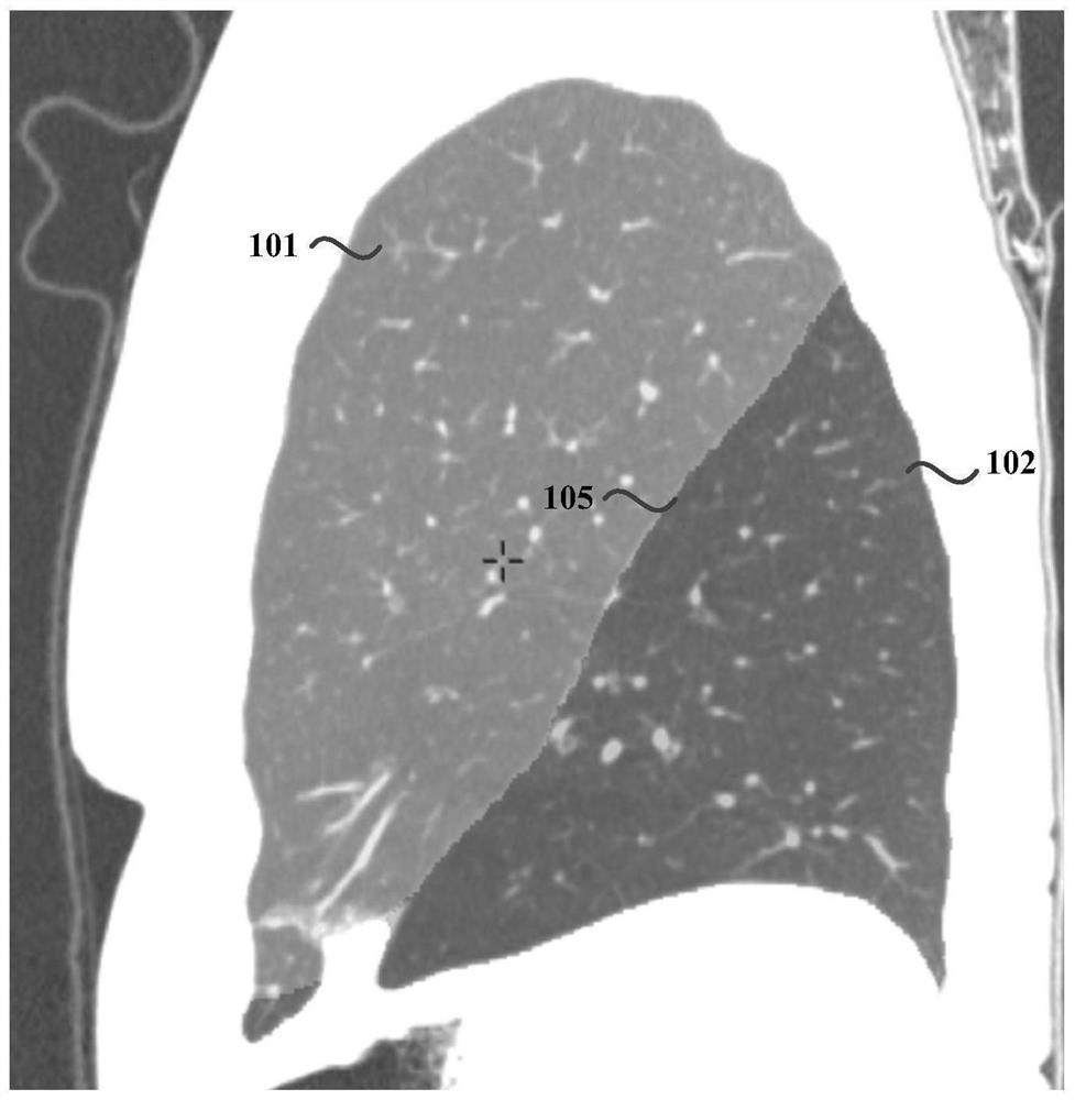
[0071] Wherein, the original lung image is a three-dimensional CT image of the lung. Fissures are the boundaries of the lung lobes that divide the complete lung into different lobar sections. The left lung of the human body contains a left oblique fissure, which divides the left lung into the left upper l...

Embodiment 2

[0096] figure 2 It is a structural diagram of a lung lobe segmentation device provided in Embodiment 2 of the present invention, and the device is suitable for processing lung medical images. The apparatus of this embodiment can be realized by means of hardware and / or software, and can generally be integrated into computer equipment or a medical image workstation.

[0097] Such as figure 2 As shown, the lung lobe segmentation apparatus specifically includes: a first segmentation module 210 , a target lung fissure acquisition module 220 and a second segmentation module 230 .

[0098] Wherein, the first segmentation module 210 is configured to perform image segmentation on the acquired original lung image based on a preset image segmentation algorithm to obtain a preliminary lung lobe segmentation image, and the preliminary lung lobe segmentation image includes a preliminary segmented lung lobe and an initial pulmonary fissure; The target lung fissure acquisition module 220 ...

Embodiment 3

[0121] image 3 It is a schematic structural diagram of the computer device in Embodiment 3 of the present invention. image 3 A block diagram of an exemplary computer device 312 suitable for use in implementing embodiments of the invention is shown. image 3 The computer device 312 shown is only an example, and should not impose any limitation on the functions and scope of use of the embodiments of the present invention.

[0122] Such as image 3 As shown, computer device 312 takes the form of a general-purpose computing device. Components of computer device 312 may include, but are not limited to: one or more processors or processing units 316 , system memory 328 , bus 318 connecting various system components including system memory 328 and processing unit 316 .

[0123] Bus 318 represents one or more of several types of bus structures, including a memory bus or memory controller, a peripheral bus, an accelerated graphics port, a processor, or a local bus using any of a v...


PUM


Abstract

The embodiment of the invention discloses a lung lobe segmentation method, device, computer equipment and storage medium. Wherein, the method includes: performing image segmentation on the collected original lung image based on a preset image segmentation algorithm to obtain a preliminary lung lobe segmentation image, the preliminary lung lobe segmentation image includes a preliminary lung lobe segmentation and an initial lung fissure; Or at least one first target lung fissure line input from the original lung image, wherein the first target lung fissure line is a complete lung fissure line or a locally adjusted lung fissure line; The split is adjusted to obtain the segmented image of the target lung lobe. The embodiment of the present invention solves the problem that the accuracy of the automatic lung lobe segmentation results is limited by the quality of lung CT image data and the accuracy of the lung lobe automatic segmentation algorithm, and cannot meet the clinical needs. The accuracy of segmentation results meets the needs of clinical diagnosis.

Description

technical field [0001] Embodiments of the present invention relate to medical image processing technology, and in particular to a lung lobe segmentation method, device, computer equipment and storage medium. Background technique [0002] The lung is the most important respiratory organ of the human body. It is located inside the chest cavity and above the diaphragm. The human lung includes five different anatomical structures, namely the lung lobes, and the lung fissure is the boundary between different lung lobes. The left lung is divided into upper and lower lobes by an oblique fissure, and the right lung is divided into upper, middle, and lower lobes by an oblique and a horizontal fissure. [0003] With the development of modern computer technology and medical imaging technology, in the diagnosis of lung diseases such as emphysema and lung cancer, higher requirements are put forward for the location of lesions, so as to provide more effective reference for lung diagnosis ...

Claims


Application Information

Patent Timeline
Patent Type & Authority Patents(China)
IPC IPC(8): G06T7/11G06T15/04
CPCG06T7/11G06T15/04G06T2207/30061G06T2207/10081
Inventor 黄石泉
Owner SHANGHAI UNITED IMAGING HEALTHCARE
Why Eureka
Try Eureka