Method for separating single cells from sample
A single-cell and sample technology, applied in the field of single-cell separation, can solve the problems of low single-cell yield and cell viability, and achieve the effect of promoting further research, high-yield and high-viability extraction
- Summary
- Abstract
- Description
- Claims
- Application Information
AI Technical Summary
Problems solved by technology
Method used
Image

Examples
Embodiment approach
[0034] According to a specific embodiment of the present invention, the method includes the following steps:
[0035] 1) In the tissue preservation solution, use sterile ophthalmic scissors to trim off the fiber, fat and necrotic tissue on the normal tissue sample;
[0036] 2) washing with sterile PBS;
[0037] 3) Put the sample into a sterile centrifuge tube, add the first part of the digestion solution (relative to the volume of 1cm 3 For normal tissue samples, the amount of the first part of the digestive solution can be 400-1000μL);
[0038] 4) Use sterile ophthalmic scissors to cut the tissue into debris (approximately 2×2×1mm 3 ), operated on ice, and the shearing time was not more than 5 minutes;
[0039] 5) Transfer the tissue debris to a sterile petri dish, add the second part of the digestive solution (relative to the volume of 1cm 3 For normal tissue samples, the amount of the second part of the digestive solution can be 5-20mL);
[0040] 6) Use a sterile Buchn...
Embodiment 1
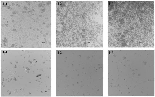
[0057] This example is used to illustrate the method for isolating single cells from normal pancreatic tissue (normal pancreas at the tumor site) of the present invention, wherein the tissue preservation solution, digestion solution, lysate and washing solution used are shown in Table 1, The enzyme activity unit value and concentration of each component in each liter of digestive juice are shown in Table 2, and the preparation method of the digestive juice is: mix type VIII collagenase, Dispase II and trypsin inhibitor, use solvent to dissolve the mixed powder, and then add DNaseI and mix well.
[0058] The samples were divided into five groups for experimentation (see Table 3), and the specific operation steps were as follows:
[0059] 1) In the tissue preservation solution, use sterile ophthalmic scissors to trim off the fiber, fat and necrotic tissue on the specimen (the normal pancreatic tissue size is about 1.5×1.5×0.5cm 3 , yellow in color, medium to soft in texture, no...
Embodiment 2
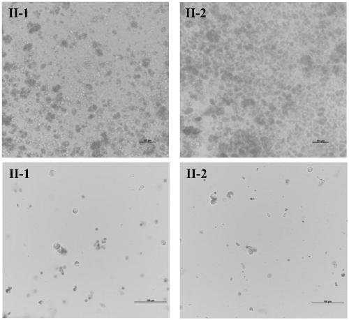
[0081] This example is used to illustrate the method of the present invention for isolating single cells from pancreatic-derived tumor tissue, wherein the tissue preservation solution, digestion solution, lysate and washing solution used are as shown in Table 1, and each liter of digestion solution (preparation method The enzyme activity unit value and concentration of each component in Example 1) are as shown in I-1 of Table 2.
[0082] 1) In the tissue preservation solution, use sterile ophthalmic scissors to trim off the pancreatic tumor sample (1.5×1.5×0.5cm 3 , Obtained by surgical resection, the patient has signed the informed consent) on the fiber, fat and necrotic tissue, etc.
[0083] 2) Wash 3 times with sterile PBS.
[0084] 3) Put the sample into a 5 mL sterile EP tube (eppendorf), and add about 500 μl of digestion solution.
[0085] 4) Use sterile ophthalmic scissors to cut the tissue into debris (approximately 2×2×1mm 3 ), operated on ice, and the shearing tim...
PUM
 Abstract
Description
Claims
Application Information
 - R&D
- Intellectual Property
- Life Sciences
- Materials
- Tech Scout
- Unparalleled Data Quality
- Higher Quality Content
- 60% Fewer Hallucinations
Browse by: Latest US Patents, China's latest patents, Technical Efficacy Thesaurus, Application Domain, Technology Topic, Popular Technical Reports.
© 2025 PatSnap. All rights reserved.Legal|Privacy policy|Modern Slavery Act Transparency Statement|Sitemap|About US| Contact US: help@patsnap.com