Transplant and method for repairing articular cartilage defects

A technology for repairing joints and articular cartilage, which is applied in the fields of medicine and biomedical engineering, and can solve the problems of side injury in the patient's donor site, difficult repair and reconstruction of large articular cartilage damage, etc., and achieve minimally invasive effects.

Inactive Publication Date: 2019-06-18
SHANGHAI SIXTH PEOPLES HOSPITAL
View PDF12 Cites 3 Cited by
  • Summary
  • Abstract
  • Description
  • Claims
  • Application Information

AI Technical Summary

Problems solved by technology

At present, costal cartilage transplantation has been routinely used in the reconstruction of the external ear and trachea, and the costal cartilage junction is used for the repair and reconstruction of interphalangeal joints, metacarpophalangeal joints, mandibular joints, elbow joints and wrist cartilage injuries. However, a rib There is only one osteochondral junction, and repairing a large area of ​​osteochondral defect with mosaic stitching of multiple rib cartilage junctions will cause a lot of side damage to the donor site, so this method is difficult to apply to large-scale cartilage damage in large joints In terms of restoration and reconstruction, there are no related literature reports

Method used


Image

  • Transplant and method for repairing articular cartilage defects
  • Transplant and method for repairing articular cartilage defects
  • Transplant and method for repairing articular cartilage defects

Examples


Embodiment 1

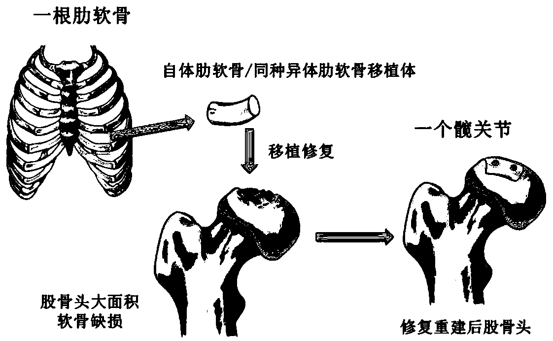
[0063] Present embodiment is the method for repairing femoral head cartilage injury with autologous costal cartilage transplantation, and its steps are as follows: figure 1 shown, including:

[0064] Under general anesthesia, the patient was placed in a supine position, and the Smith-Peterson approach was used to incise the skin, subcutaneous tissue, and fascia lata in sequence, paying attention to protecting the anterolateral femoral cutaneous nerve, fully exposing the anterior joint capsule of the hip joint, and paralleling a "T" shape cut open. After the hip joint is fully released, the femur is extremely adducted and externally rotated to disengage the femoral head. Thoroughly clean the degenerated and necrotic bone and cartilage tissue of the femoral head, and confirm the osteochondral defect area on the surface of the femoral head after cleaning. According to the bone defect, the ipsilateral iliac bone fragment was taken in the same incision to reconstruct the subchond...

Embodiment 2

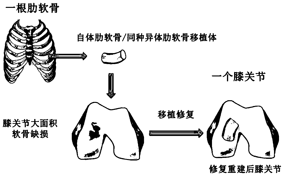
[0066] This embodiment is a method for autologous costal cartilage transplantation to repair knee articular cartilage damage, and its steps are as follows: figure 2 shown, including:

[0067] After the anesthesia takes effect, the patient is placed in the supine position. Routine disinfection, sterile towels, and tourniquets on the affected limbs to reduce bleeding. The left knee parapatellar medial longitudinal incision skin, superficial deep fascia, incised medial patellar support ligament, turned the patella laterally to expose the damaged cartilage surface of the medial femoral condyle. Thoroughly remove diseased cartilage and subchondral bone tissue until fresh bleeding occurs on the bone bed. The ipsilateral ilium was harvested and transplanted to reconstruct the subchondral bone defect. At the same time as the knee surgery, another group of doctors stood on the right side of the patient to harvest the costal cartilage. The skin was incised about 4 cm along the long...

Embodiment 3

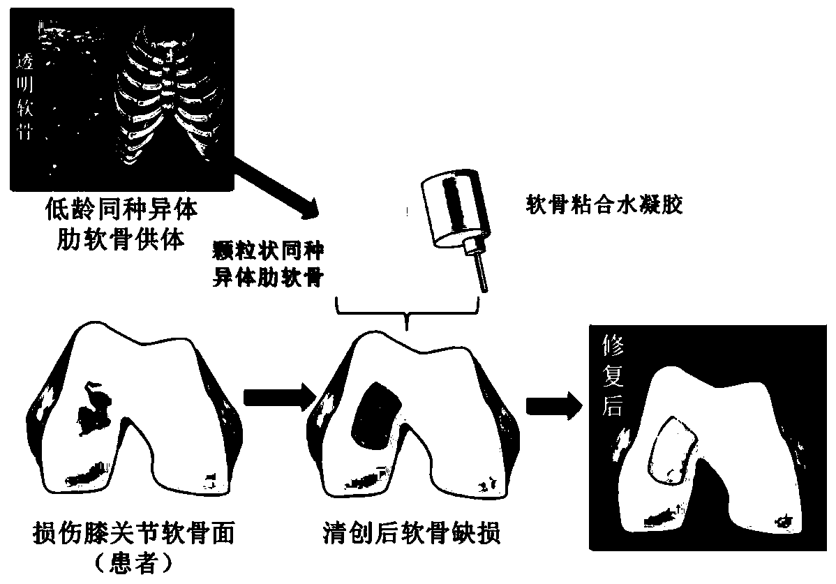
[0069] This embodiment is a method for repairing femoral head cartilage damage with allogeneic costal cartilage transplantation. Specifically include:

[0070] Collect and preserve allogeneic cartilage: take the costal cartilage from a young donor and place it in a preservation solution (serum-free medium and pH buffer), and remove the preservation solution before use;

[0071] Under general anesthesia, the patient was placed in a supine position, and the Smith-Peterson approach was used to incise the skin, subcutaneous tissue, and fascia lata in sequence, paying attention to protecting the anterolateral femoral cutaneous nerve, fully exposing the anterior joint capsule of the hip joint, and paralleling a "T" shape cut open. After the hip joint is fully released, the femur is extremely adducted and externally rotated to disengage the femoral head. Thoroughly clean the degenerated and necrotic bone and cartilage tissue of the femoral head, and confirm the osteochondral defect...


PUM

PropertyMeasurementUnit
sizeaaaaaaaaaa

Abstract

The invention provides a transplant for repairing articular cartilage defects. The transplant comprises at least one of autologous costicartilage and allogeneic costicartilage; the transplant can be specially a whole-section costicartilage or cartilage particle combined hydrogel transplant. The invention further relates to application of the transplant and a method for repairing the articular cartilage defects. The transplant and method for repairing the articular cartilage defects and the application of the transplant have the advantages that by adopting costal cartilage grafting, vice-damageis small, and minimally invasive operation can be carried out to avoid the risk of prosthesis loosening, infection and other complications which are caused by artificial joint replacement; the amountof the costicartilage is sufficient, and the needs of multiple times of cartilage reconstruction and revision surgeries can be met; it can be achieved that the reconstruction of the damaged articularcartilage surface is personalized, and the repairing method with higher safety and stronger operability is provided for patients with the articular cartilage defects.

Description

technical field [0001] The invention relates to the technical fields of medicine and biomedical engineering, in particular to an implant and a method for repairing articular cartilage defects. Background technique [0002] Articular cartilage is hyaline cartilage, mainly composed of chondrocytes, cartilage matrix, and type II collagen. It has complex biomechanical characteristics and high durability, but its self-repair ability is limited, and defects caused by natural degeneration or trauma will cause irreversible damage to its structure and function. [0003] Because the service life of artificial joints is limited, for young and middle-aged people, the cartilage of the hip and knee joints is damaged. Once artificial joint replacement is performed, it means multiple surgical revisions. Therefore, when trauma, necrosis, infection and other factors lead to articular surface cartilage degeneration and injury, artificial joint replacement should be avoided as much as possible...

Claims


Application Information

Patent Timeline
Patent Type & Authority Applications(China)
IPC IPC(8): A61F2/28A61F2/30
CPCA61F2/30756A61F2002/30062A61F2310/00371A61F2310/00377A61F2310/00383A61L27/3612A61L27/3654A61L27/52A61L2430/06A61F2002/30764A61F2210/0004A61L27/24A61L27/3817A61L27/54
Inventor 张长青杜大江
Owner SHANGHAI SIXTH PEOPLES HOSPITAL
Why Eureka
Try Eureka