Supercharge Your Innovation With Domain-Expert AI Agents!

Detection probe and applications thereof

A technology for detecting probes and probes, which is used in the determination/inspection of microorganisms, biochemical equipment and methods, DNA/RNA fragments, etc. Laboratory conditions require low effects

Pending Publication Date: 2019-07-02
SHENZHEN INST OF ADVANCED TECH
View PDF8 Cites 1 Cited by
  • Summary
  • Abstract
  • Description
  • Claims
  • Application Information

AI Technical Summary

Problems solved by technology

[0005] CN105807064A provides a luciferase complementary quantum dot biosensor and its construction method and application thereof. The luciferase complementary quantum dot biosensor includes quantum dots, luciferase amino-terminal fragments, luciferase carboxyl-terminal fragments, A probe that specifically recognizes the analyte and a substrate that can undergo a bioluminescence reaction with the luciferase; this invention combines the optical advantages of the quantum dot sensor, by realizing the amino-terminal fragment and the carboxyl-terminal fragment of the luciferase on the surface of the quantum dot Induced complementarity, reconstruction of catalytic function, construction of a new type of high-sensitivity biosensor, which can be applied to the high-target detection of various biomarkers, and is suitable for accurate quantification of target detection substances in a homogeneous system, but the method steps complicated and time-consuming

Method used


Image

  • Detection probe and applications thereof
  • Detection probe and applications thereof
  • Detection probe and applications thereof

Examples


Embodiment 1

[0054] Example 1 Assembly Kit

[0055] Probe A and probe B were designed according to the target series miR208a, wherein the 5′ end of probe A was modified with Gaussia luciferase, the 3′ end of probe B was modified by quantum dot QD655, the target nucleic acid, probe A and probe The sequence of B is shown in Table 1:

[0056] Table 1

[0057]

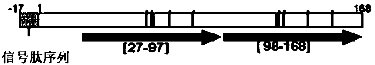
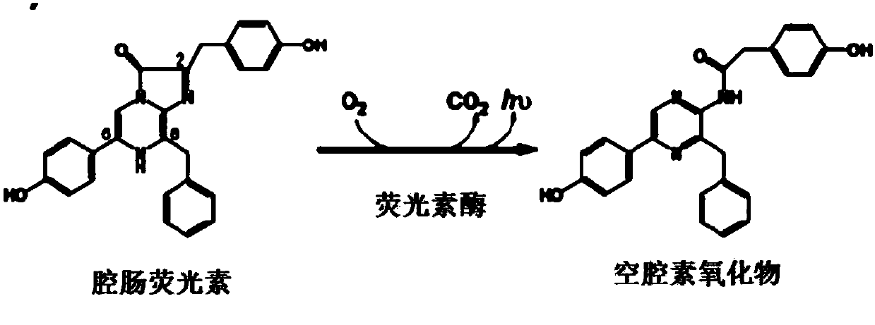
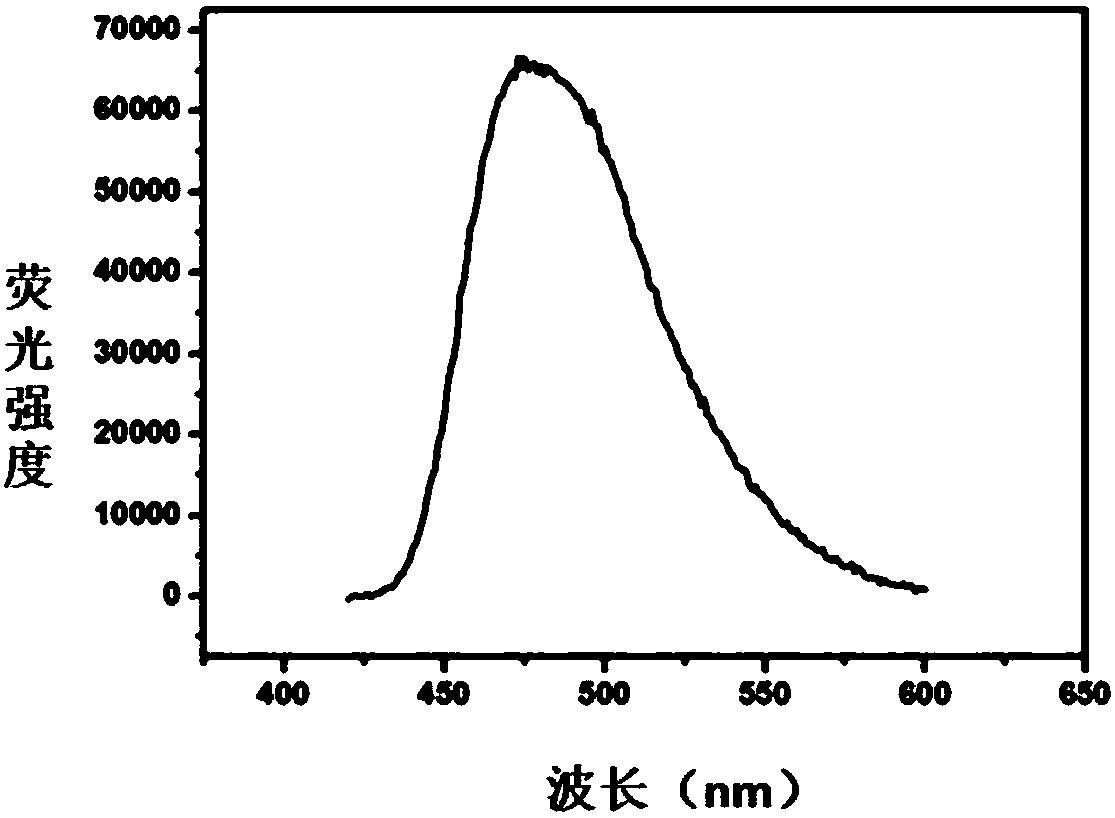
[0058] The primary structure distribution of Gaussia luciferase is as follows figure 1 As shown, the amino acid sequence 27-97 (hGL-27-97) and 98-168 (hGL-98 / 168) constitute two independent enzyme catalytic structures; the bioluminescent schematic diagram of Gaussia luciferase is shown in figure 2 Shown; Gaussia luciferase emission spectrum characteristic figure is shown in image 3 shown.

[0059] Other common reagents include: 50 mM Tris-HCl buffer pH=7.4.

[0060] Probe A and probe B at a concentration of 100 nM, luciferase substrate at a concentration of 0.5 μg / μL, magnesium ion solution at a concentration of 10 mM, other ...

Embodiment 2

[0061] Embodiment 2 experimental detection

[0062] (1) adding probe A and probe B with a concentration of 100 nM and luciferase substrate with a concentration of 0.5 μg / μL to a magnesium ion solution with a concentration of 10 nM to obtain a mixed solution;

[0063] (2) adding the nucleic acid to be tested to the mixture obtained in step (1), and performing a hybridization reaction at 37° C. for 30 minutes;

[0064] (3) Detect the fluorescence intensity after the hybridization reaction, and calculate the fluorescence intensity ratio between the luciferase and the quantum dot;

[0065] The sequence of the nucleic acid to be tested miR208b is shown in SEQ ID NO: 4:

[0066] SEQ ID NO:4AUA AGA CGA ACA AAA GGU UUG U

Embodiment 3

[0068] Compared with Example 2, except that the nucleic acid to be tested is changed to miR-155, other conditions are the same as Example 2;

[0069] The sequence of MiR-155 is shown in SEQ ID NO:5:

[0070] SEQ ID NO:5UUA AUG CUA AUC GUG AUAGGG GU


PUM


Abstract

The invention provides a detection probe and applications thereof. The probe comprises a probe A labeled with luciferase and a probe B labeled with quantum dots; partial sequences of one end of the probe A and one end of the target nucleic acid are complementary, partial sequences of one end of the probe B and the other end of the target nucleic acid are complementary, and partial sequences of theother end of the probe A and the other end of the probe B are complementary; and the probe A, the probe B and the target nucleic acid are hybridized to form a stable T-shaped structure, and the luciferase and the quantum dots can be mutually paired to generate bioluminescence resonance energy transfer. By designing the specific probes, the invention skillfully enables the probes and the nucleic acid to form a stable T structure. The reaction conditions are optimized by utilizing the principle of bioluminescence energy resonance transfer, single base mutation can be accurately distinguished, the target nucleic acid can be effectively detected, the operation is simple, the required time is short, convenience and rapidness are realized, and the cost is low.

Description

technical field [0001] The invention relates to the field of biotechnology, in particular to a detection probe and its application. Background technique [0002] Bioluminescence resonance energy transfer (BRET) is a non-radiative energy transfer between a bioluminescent protein such as luciferase (donor) and a fluorescent substance (acceptor). Because BRET does not require exogenous excitation, low background, and high sensitivity, it has been widely used as a micro-distance optical ruler in the fields of protein interaction, live imaging, nucleic acid analysis, protease detection, and high-throughput screening. BRET technology is based on the phenomenon of resonance energy transfer in some marine animals (such as Renilla luciferase) and results in a non-radioactive energy transfer between an energy donor and an energy acceptor. The energy donor in BRET is a luminescent luciferase, which emits a spectrum of the corresponding wavelength in the presence of the corresponding s...

Claims


Application Information

Patent Timeline
IPC IPC(8): C12Q1/6818C12Q1/66C12N15/11
CPCC12Q1/6818C12Q1/66C12Q2563/107C12Q2565/101
Inventor 金宗文罗擎颖刘琳
Owner SHENZHEN INST OF ADVANCED TECH
Features
  • R&D
  • Intellectual Property
  • Life Sciences
  • Materials
  • Tech Scout
  • Unparalleled Data Quality
  • Higher Quality Content
  • 60% Fewer Hallucinations
Learn More