
Micro-fluidic chip for constructing brain function unit model and construction method

A technology of microfluidic chips and construction methods, applied in artificial cell constructs, biochemical equipment and methods, microorganisms, etc., to achieve the effects of strong versatility, weak in vitro proliferation ability, and easy regulation

Active Publication Date: 2019-08-09
FIRST AFFILIATED HOSPITAL OF DALIAN MEDICAL UNIV
View PDF17 Cites 6 Cited by
  • Summary
  • Abstract
  • Description
  • Claims
  • Application Information

AI Technical Summary

Problems solved by technology

However, this technique requires precise regulation of the supply of oxygen and nutrients to best approximate the true state of the cerebrospinal fluid

Method used


Image

  • Micro-fluidic chip for constructing brain function unit model and construction method
  • Micro-fluidic chip for constructing brain function unit model and construction method
  • Micro-fluidic chip for constructing brain function unit model and construction method

Examples


Embodiment 1

[0035] Using an assembled and integrated microfluidic device, the in vitro neural-vascular unit is constructed to simulate the functional unit of the brain.

[0036] Design and preparation of functionalized microfluidic chips that mimic brain functional units:

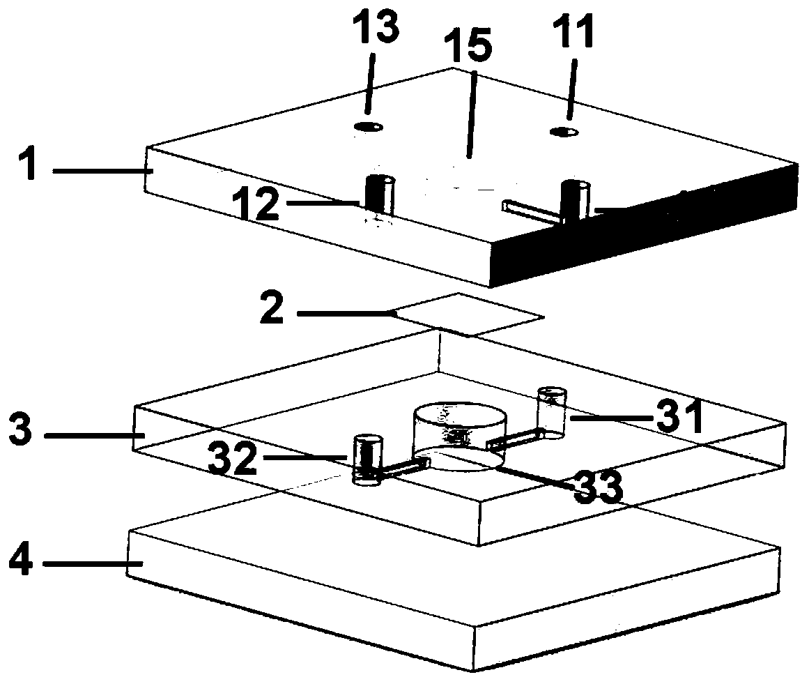
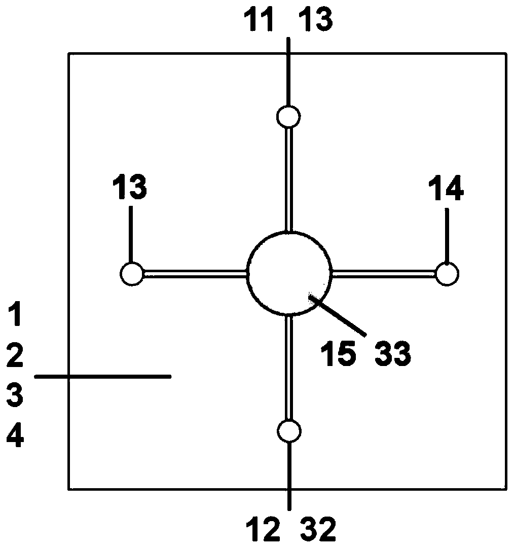
[0037] 1) Devices for functionalized microfluidic chips for simulating brain functional units, such as figure 1 , 2 shown. The whole device is composed of the first elastic layer 1 , the middle layer 2 , the second elastic layer 3 and the substrate 4 sequentially from the upper layer to the bottom layer. The first elastic layer 1 and the second elastic layer 3 are respectively PDMS layers, the intermediate layer 2 is a polycarbonate film, and the substrate 4 is a glass substrate.

[0038] Wherein, the first elastic layer 1 is provided with a first liquid inlet 11, a first liquid outlet 12, a second liquid inlet 13, a second liquid outlet 14 and a first culture chamber 15 through it, the The first culture chamber 15...

Embodiment 2

[0047] Using microfluidic chips to construct models of brain functional units:

[0048] (1) Culture and identification of human brain microvascular endothelial cells and neural stem cells:

[0049] 1) Culture and identification of brain microvascular endothelial cells.

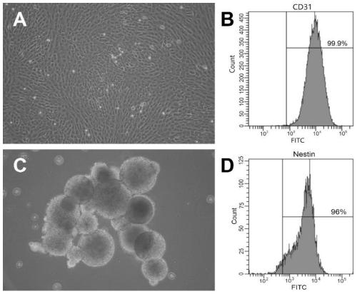
[0050] Human brain microvascular endothelial cells were cultured in DMEM medium containing 10% fetal bovine serum, and grown as a single layer adherently. After 90% of the cells were confluent, they were trypsinized and passaged. For phenotype identification, adherent cells were digested with trypsin to form a single-cell suspension, washed with PBS, then added with FITC-labeled CD31 antibody, and incubated in the dark. After washing with PBS, the cells were detected by flow cytometry. Cerebral microvascular endothelial cells image 3 As shown in A, the quantitative characterization by flow cytometry is as follows image 3 As shown in B, the positive rate of CD31 reached 99.9%.

[0051] 2) Primary extract...

Embodiment 3

[0060] Identification of the survival of each cell component in the brain functional unit model prepared in Example 2:

[0061] 1) Identification of microvascular endothelial cell activity in the brain functional unit model.

[0062] After 7 days of perfusion culture, the two PDMS layers were disassembled, and the polycarbonate membrane attached to the microvascular endothelial cells was carefully taken out and placed in a small dish. After washing with PBS, Calcein and PI dyes were added, and incubated in the dark. The dye was discarded and detected under a confocal fluorescence microscope after adding PBS. The sample was scanned in 20 slices, and the scanned images of 5 slices at equal intervals were taken, and the number of Calcein-expressing cells and PI-expressing cells were counted, and the percentage of living cells was calculated. The morphology of brain microvascular endothelial cells on the polycarbonate membrane is as follows: Figure 4 As shown in A, the survival...


PUM

PropertyMeasurementUnit
heightaaaaaaaaaa
heightaaaaaaaaaa
pore sizeaaaaaaaaaa

Abstract

The invention provides a micro-fluidic chip for constructing a brain function unit model and a construction method thereof. The micro-fluidic chip includes a first elastic layer, a second elastic layer, a middle layer and a substrate, wherein the first elastic layer is provided with a first culture chamber, a liquid inlet and a liquid outlet, the second elastic layer is provided with a second culture chamber, a liquid inlet and a liquid outlet, the middle layer is located between the first elastic layer and the second elastic layer, and the substrate fits the bottom of the second elastic layer. First cells such as microvascular endothelial cells attached to the middle layer are cultured in the first culture chamber, second cells such as primary neural stem cells are cultured in the secondculture chamber, the liquid inlets and the liquid outlets are connected with the corresponding culture chambers through micro-channels to form a perfusion culture channel, and thus the purpose of using a relatively simple method and least cell types for simulating and constructing a relatively complex brain structure functional unit is achieved. The micro-fluidic chip for constructing the brain function unit model and the construction method thereof can be used for constructing models of various neural system diseases, evaluating the safety and efficacy of drugs and conducting dosage screeningand the like, so that a good carrier and technical support are provided for clinical drug screening.

Description

technical field [0001] The invention belongs to the research field of tissue engineering-in vitro construction of tissue functional units, and in particular relates to a microfluidic chip and a construction method for constructing brain functional unit models. Background technique [0002] Nerve-vascular unit, that is, a complex composed of endothelial cells and extracellular matrix, astrocytes, pericytes, neurons and their axons, and other supporting cells (microglia, oligodendrocytes), etc. The body is the basic unit of structure and function of the nervous system. These components are in close contact and coordinated with each other to form a complete anatomical and functional unit, efficiently and precisely regulate cerebral blood flow, and jointly maintain the homeostasis of the internal environment of brain tissue. The concept of nerve-vascular unit is proposed to emphasize the importance of the interconnection and mutual influence between neurons, glial cells and cer...

Claims


Application Information

Patent Timeline
Patent Type & Authority Applications(China)
IPC IPC(8): C12M3/00C12N5/0735C12N5/074C12N5/0775C12N5/0797C12N5/071
CPCC12M23/16C12M23/34C12N5/069C12N5/0691C12N5/0623C12N5/0662C12N5/0696C12N5/0606C12N2513/00
Inventor 刘晶魏文娟马静云陈维功
Owner FIRST AFFILIATED HOSPITAL OF DALIAN MEDICAL UNIV
Learn More