Intra-cellular miRNA quantifying method based on unimolecule fluorescence imaging

A single-molecule imaging, cell technology, applied in fluorescence/phosphorescence, biochemical equipment and methods, microbial assay/inspection, etc., can solve the problems of difficult miRNA quantification, many interfering substances, false positive signals, etc.

Inactive Publication Date: 2019-09-13
BEIJING UNIV OF CHEM TECH
View PDF3 Cites 1 Cited by
  • Summary
  • Abstract
  • Description
  • Claims
  • Application Information

AI Technical Summary

Problems solved by technology

The second method is currently widely used. The imaging method is mainly fluorescent imaging, and other methods such as Raman imaging are also included. However, the common imaging mode can only obtain the relative expression level by comparing the signal intensity per unit area, and it is dif...

Method used


Image

  • Intra-cellular miRNA quantifying method based on unimolecule fluorescence imaging
  • Intra-cellular miRNA quantifying method based on unimolecule fluorescence imaging
  • Intra-cellular miRNA quantifying method based on unimolecule fluorescence imaging

Examples


Embodiment 1

[0044] Example 1. Using single-molecule fluorescence imaging to detect miR-21 in A549 cells

[0045] (1) Cell fixation

[0046] Use a pipette gun to add 200 μL of 4% paraformaldehyde solution to the confocal culture dish (A549 cells and Hela cells inoculated from the culture bottle into the confocal culture dish) to make it cover the surface of the culture dish and place it on the cell surface. In the incubator, fix for 20-30min; remove the paraformaldehyde solution with a pipette gun, and wash the cells 3 times with 1×PBS;

[0047] Preparation of 4% paraformaldehyde solution: add 40 grams of paraformaldehyde to 800 ml 1×PBS, heat to 60° C., and keep stirring until transparent; adjust the pH to 7.3 with NaOH solution; then adjust the volume to 1000 ml.

[0048] (2) Nuclear dye staining

[0049] Use a pipette gun to draw 1 μL of 1mg / mL DAPI stain and add it to 199 μL of 1×PBS, mix well, and then use a pipette gun to transfer to a confocal culture dish so that it covers the su...


PUM


Abstract

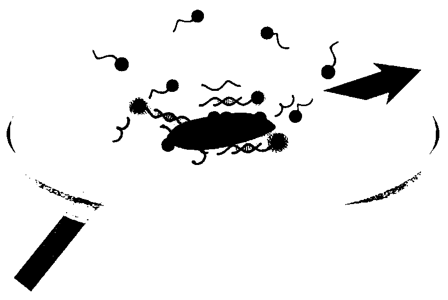
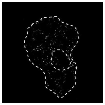
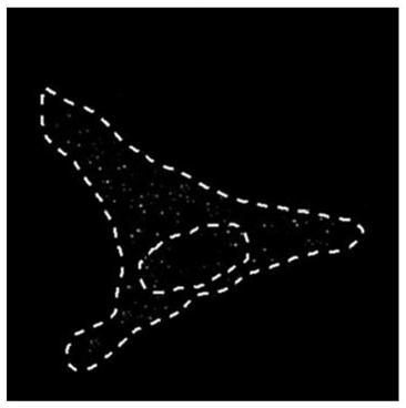
The invention discloses an intra-cellular miRNA quantifying method. The method comprises the following steps of (1) fixing excised cells, and then performing cell nucleus dyeing; (2) adding a buffer solution containing fluorescent probes to the cells, and enabling the fluorescent probes and intra-cellular miRNA to be detected to generate reversible combination; (3) placing the cells on an objective table of a TIRF microscope, performing detection, recording the shape of the cells at a bright field, performing fluorescence recording of the position of a cell nuclei at a wide field, and recording unimolecule fluorescence intensity-time track in an HILO mode; and (4) performing signal filtration to obtain a unimolecule imaging graph in which the unimolecule fluorescence intensity-time track presenting ON-OFF alternate changes, namely an intra-cellular miRNA unimolecule imaging graph, and performing counting so as to obtain the quantity of intra-cellular miRNA. According to the method disclosed by the invention, an "in-situ quantifying imaging" manner is provided, high-identification degree unimolecule dynamics fingerprint signals are used, unimolecule positioning and pinpoint countingare performed on the intra-cellular miRNA, and the technical bottleneck that intra-cellular miRNA quantifying depends on RNA extraction is broken through.

Description

technical field [0001] The invention relates to an miRNA cell imaging method, in particular to an intracellular miRNA quantification method based on single-molecule fluorescence imaging, and belongs to the field of object imaging and detection. Background technique [0002] microRNA (miRNA) is a non-coding RNA with 22 nucleotides. miRNA regulates gene expression by binding to mRNA and is involved in almost all cellular pathways, including differentiation, metabolism, apoptosis, signal transduction, etc. A number of pre-clinical studies have shown that the contents of various miRNAs in tumor cells are different from those in healthy people, and their contents are also different in different stages of tumor development. miRNA is expected to become a new generation of early diagnostic markers for tumors. The development of reliable intracellular miRNA detection methods to achieve accurate quantification of intracellular miRNA can, on the one hand, accelerate miRNAs to become s...

Claims


Application Information

Patent Timeline
IPC IPC(8): C12Q1/68G01N21/64
CPCC12Q1/68G01N21/6486
Inventor 苏昕李丽娜邓莹楠王丛珊付胜男
Owner BEIJING UNIV OF CHEM TECH
Why Eureka
Try Eureka