
Retinitis pigmentosa mutant site and application thereof

A retinal pigment and mutation site technology, applied in the field of 35 retinitis pigmentosa mutation sites, can solve the problems of unclear pathogenicity, increased birth rate of patients, and incomplete family information

Pending Publication Date: 2019-09-17
EYE & ENT HOSPITAL SHANGHAI MEDICAL SCHOOL FUDAN UNIV
View PDF6 Cites 6 Cited by
  • Summary
  • Abstract
  • Description
  • Claims
  • Application Information

AI Technical Summary

Problems solved by technology

However, like all medical interventions, genetic testing has some specific risks
For example, a problem often encountered in the current detection process is the judgment of pathogenicity of multiple mutation sites. Even though there are strict pathogenicity judgment standards, such as the ACMG standard of the American Medical Genetics Association, they are limited by various factors (such as : Incomplete family information, unreported loci, gene heterogeneity, etc.), the pathogenicity of most loci still cannot be clarified in clinical work
For example, a false positive result from a genetic test can affect a patient's family planning, create feelings of anxiety or guilt, and can even disrupt a patient's relationships with other family members
False negative results of genetic testing delay the diagnosis and treatment of patients, increase the birth rate of patients, etc.

Method used


Image

  • Retinitis pigmentosa mutant site and application thereof
  • Retinitis pigmentosa mutant site and application thereof
  • Retinitis pigmentosa mutant site and application thereof

Examples


Embodiment 1

[0037] Example 1: Gene mutation detection in a four-generation family with retinitis pigmentosa (RP)

[0038] 1. Experimental method

[0039] 1. Collection of clinical resources of the family and establishment of a genetic resource bank

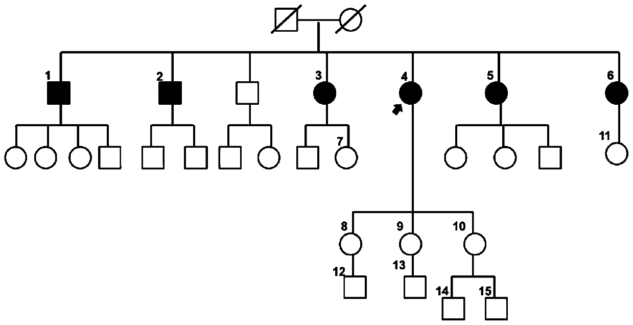
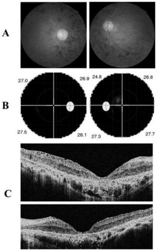
[0040] The clinical data and blood samples of each member of the family were collected, see the family diagram figure 1 . Clinical data mainly include personal medical history, family history, best corrected visual acuities (BCVAs), slit lamp examination, fundus photography, color vision examination, visual field examination (Humphrey perimetry), visual evoked potentials (visual evoked potentials; VEP) ), full field electrophysiology (electroretinography; ERG), fundus fluorescein angiography (FFA) and optical coherence tomography (optical coherence tomography; OCT), etc. Peripheral blood was extracted from patients and family members, and DNA was extracted with a blood genome DNA extraction kit (Qiagen, Hilden, Germany).

[0041] 2. Detec...

Embodiment 2

[0079] Example 2: Verification in the RP population for the two pathogenic sites detected in Example 1

[0080] 1. Experimental method

[0081] 1. RP Patient Collection

[0082] A comprehensive ophthalmic examination was performed on patients with suspected RP, and clinical resources were collected and a genetic resource bank was established for patients according to the method in Case 1.

[0083] 2. Perform high-throughput next-generation sequencing on the patient according to the method in Example 1 to detect pathogenic mutations in RP patients.

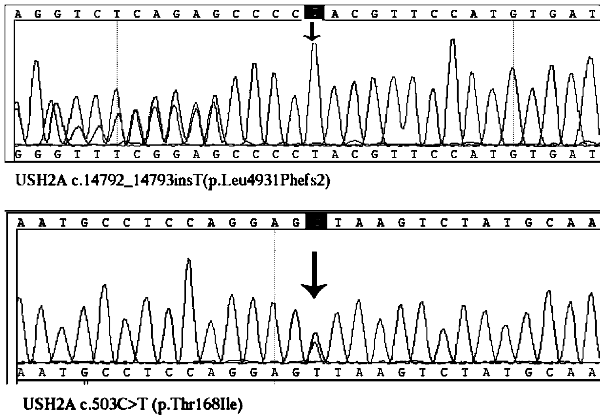
[0084] 3. Three other patients with c.14792_14793insT (p.Leu4931Phefs2) and two patients with c.503C>T (p.Thr168Ile) were detected from 1242 confirmed RP patients, and no other suspected pathogenic factors were found genetic mutation site. An example is selected below for illustration.

[0085] 4. c.14792_14793insT(p.Leu4931Phefs2) and a reported pathogenic mutation USH2A c.2802T>G p.Cys934Trp were detected in patient A, see th...


PUM


Abstract

The invention relates to a retinitis pigmentosa mutant site and an application thereof. The retinitis pigmentosa mutant site is based on clinical resources, through a gene capture chip and a deep sequencing technique, through genetics, clinical manifestation and crowd verification are used for combination, the situation that detected 35 mutant sites are retinitis pigmentosa new infective sites is successfully confirmed, a new molecular biology base is provided for disease diagnosis, and a retinitis pigmentosa diagnosis reagent kit can be developed based on the 35 mutant sites.

Description

technical field [0001] The invention belongs to the field of disease diagnosis and gene detection, and specifically relates to 35 retinitis pigmentosa mutation sites and applications thereof. Background technique [0002] Retinitis pigmentosa (RP) is a group of progressive and blinding hereditary fundus diseases characterized by damage to retinal photoreceptors (rods and cones) and pigment epithelium. The incidence rate of RP in the world is 1 / 3000-1 / 5000, and the incidence rate of the Chinese population can be as high as 1 / 1000. The main clinical features of the disease are: progressive visual field defect, night blindness, retinal bone cell-like pigmentation and abnormal electroretinogram. The age of onset varies from birth to old age. About 20%-30% of RP patients have extraocular symptoms including hearing loss, obesity, polydactyly, and neurological lesions. These extraocular abnormalities are easily overlooked or confused. Furthermore, the clinical presentation of R...

Claims


Application Information

Patent Timeline
IPC IPC(8): C12Q1/6883C12N15/11
CPCC12Q1/6883C12Q2600/156
Inventor 吴继红张圣海高凤娟徐格致胡方园孙兴怀卢奕徐萍王丹丹张道微
Owner EYE & ENT HOSPITAL SHANGHAI MEDICAL SCHOOL FUDAN UNIV
Learn More