Imaging method of RNA tailing and structures of in-situ cells

An imaging method and cell technology, applied in the direction of biochemical equipment and methods, microbial measurement/inspection, etc., can solve the problems of subcellular structure and copy number information cannot be obtained, and achieve simple reaction system, high reaction efficiency, and accurate imaging Effect

Active Publication Date: 2019-11-22
XI AN JIAOTONG UNIV
View PDF3 Cites 0 Cited by
  • Summary
  • Abstract
  • Description
  • Claims
  • Application Information

AI Technical Summary

Problems solved by technology

However, RNA subcellular structure and copy numb

Method used


Image

  • Imaging method of RNA tailing and structures of in-situ cells
  • Imaging method of RNA tailing and structures of in-situ cells
  • Imaging method of RNA tailing and structures of in-situ cells

Examples


Embodiment 1

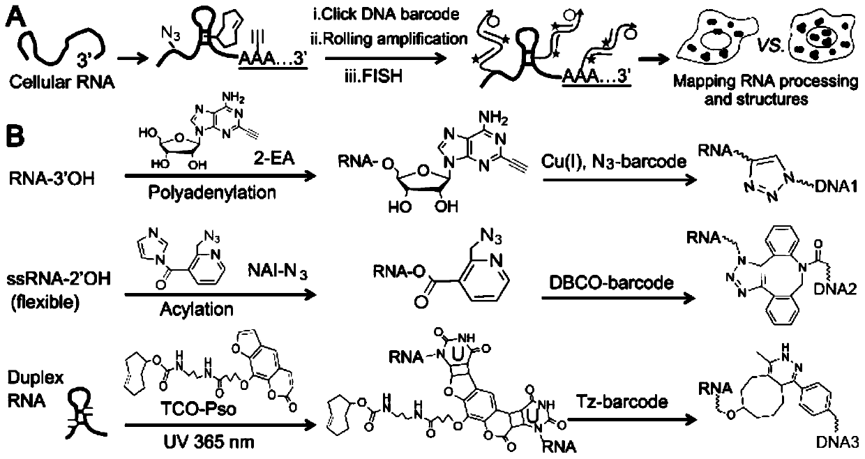
[0076] Example 1 Imaging of in situ cellular RNA tailing using the ClickerFISH method

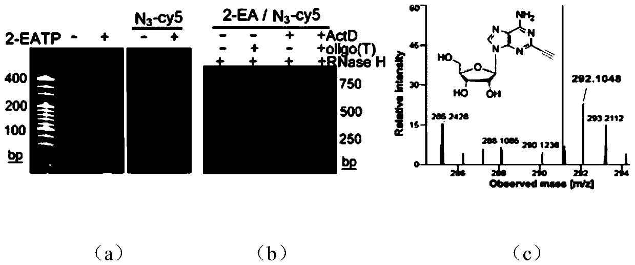
[0077] Using the MBA-MD-231 human breast cancer cell line as the basic model, live cells were cultured with 2 μM transcription inhibitor ActD for 1 hour, and continued to culture with 100 μM 2-EA for 1 hour without changing the medium; after washing the cells with PBS for 3 times, Fix the cells with 4% (mass / volume) paraformaldehyde at room temperature for 10 minutes, wash the cells 3 times with PBS, and permeabilize the cells with 0.5% (volume / volume) Triton X-100 at room temperature for 5 minutes; wash with PBS After cells 3 times, add click chemistry amplification reagents including 1 μM azide-modified priming strand, 2.5 μg / mL yeast tRNA, 1 mM CuSO 4 React with 100mM sodium ascorbate at room temperature for 1 hour; wash the cells 3 times with 1xSSC, and perform a ring amplification experiment to achieve signal amplification. The specific process is as follows: First, 20μL 1xSSC hybridiz...

Embodiment 2

[0079] Example 2 Using the ClickerFISH method to perform RNA tailing and imaging of single-strand and double-strand structures in different cell lines

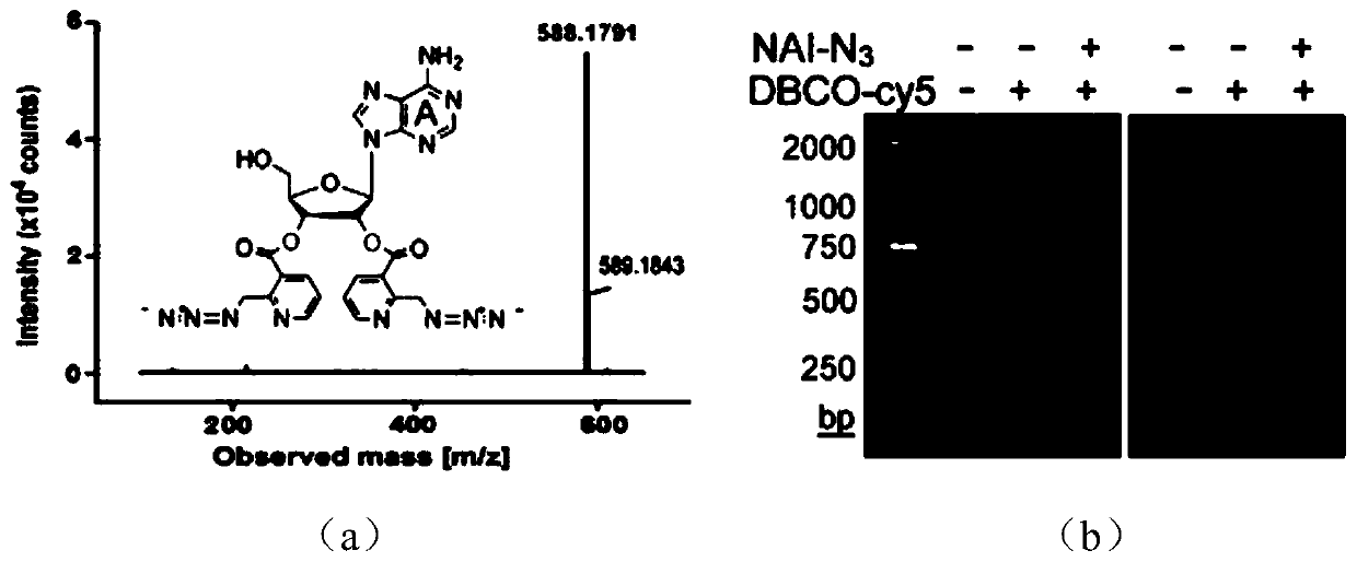
[0080]Three different cell lines of MCF-10A human normal breast epithelial cells, MCF-7 human breast cancer cells and MBA-MD-231 human breast cancer cells were selected for the following process. Live cells were cultured with 2 μM transcription inhibitor ActD for 1 hour, and continued to culture with 100 μM 2-EA for 1 hour without changing the medium; after washing the cells with PBS for 3 times, fix the cells with 4% (mass / volume) paraformaldehyde at room temperature After 10 minutes, the cells were washed 3 times with PBS, and the cells were permeabilized with 0.5% (volume / volume) Triton X-100 at room temperature for 5 minutes; after the cells were washed 3 times with PBS, the RNA single-stranded acylation reagent NAI- N3, the reaction process is: 2mMNAI-N3, 1U RNase inhibitor react with fixed cells for 30 minutes at 37°C; a...


PUM


Abstract

The invention discloses an imaging method of RNA tailing and structures of in-situ cells, and belongs to the technical field of cellular imaging. The imaging method of the RNA tailing and the structures of the in-situ cells takes intracellular RNA tailing and various structures as research targets; and then, click-encoded rolling FISH (ClickerFISH) is adopted for realizing RNA tailing as well as single-strand and double-strand structures while realizing single-cell single-molecule imaging. The reaction system utilized by the imaging method of the RNA tailing and the structures of the in-situ cells disclosed by the invention is simple, high in reaction efficiency, as well as capable of realizing rapid and accurate imaging of intracellular RNA. Being used for studying intracellular RNA tailing, it has been found that the occurrence location and the length of RNA tailing are different in different cell lines and cell cycles; and moreover, being used for studying intracellular RNA single-strand and double-strand structures, it has been found that the distribution of the single-strand and double-strand structures are different in different cell lines and cell cycles.

Description

technical field [0001] The invention belongs to the technical field of in situ cell imaging, and relates to an imaging method for in situ cell RNA tailing and structure. Background technique [0002] RNA plays an important role in many biological processes such as gene coding, non-coding, expression and regulation, and its synthesis, tailing and high-level structure lay the foundation for various biological functions. The 3′ end polyadenylation tailing, also known as poly(A) tailing, affects the stability and translation process of RNA, and is closely related to tumor activation and metastasis. In addition, due to conformational flexibility, RNA can fold into complex structures such as stem-loops, which is critical for its information transmission, regulatory and catalytic roles. Therefore, determining RNA tailing and structure can help to understand its function and network of action. However, due to the lack of identification and monitoring methods, it is difficult to ob...

Claims


Application Information

Patent Timeline
IPC IPC(8): C12Q1/6841
CPCC12Q1/6841C12Q2525/173C12Q2531/125C12Q2563/107C12Q2525/307
Inventor 赵永席陈锋白敏薛静
Owner XI AN JIAOTONG UNIV
Why Eureka
Try Eureka