Biological material taken from adipose tissue, and preparation method and application of biological material

A biomaterial, adipose tissue technology, used in tissue regeneration, oil/fat/wax inactive ingredients, drug combinations, etc., can solve problems such as unclear cell components, reduce side effects, remove immunogenicity, and high safety Effect

Pending Publication Date: 2019-11-26
上海金致投资管理有限公司
View PDF6 Cites 11 Cited by
  • Summary
  • Abstract
  • Description
  • Claims
  • Application Information

AI Technical Summary

Problems solved by technology

Because nanofat contains stromal cells and growth factors, previous studies have believed that cellular components in nanofat play an important role (

Method used


Image

  • Biological material taken from adipose tissue, and preparation method and application of biological material
  • Biological material taken from adipose tissue, and preparation method and application of biological material
  • Biological material taken from adipose tissue, and preparation method and application of biological material

Examples


Embodiment 1

[0037] Example 1 Preparation of Biomaterials from Adipose Tissue

[0038] The fat was obtained from the patient under the condition of obtaining informed consent. The fat obtained by suction or surgical resection was shredded and rinsed three times with normal saline, placed in a 50ml centrifuge tube, and placed in a centrifuge at 1,200r / rpm After centrifuging for 3 minutes, drain the excess liquid at the bottom and the uppermost layer of fat, collect the fat in the middle layer, and obtain the crude fat product. The crude fat was mechanically emulsified by two 10ml injection syringes connected with a three-way pipe at an average speed of 60 to 120 times to obtain nano fat. Then put the nano-fat into a 50ml test tube, centrifuge at 1,500 / rpm for 5 minutes, collect the transparent liquid in the middle layer, and obtain it.

[0039] Or freeze the nanofat in a -80°C refrigerator (or liquid nitrogen), then thaw in a 37°C water bath, repeat freezing and thawing once or twice, cent...

Embodiment 2

[0041] The growth factor detection that embodiment 2 biological material contains

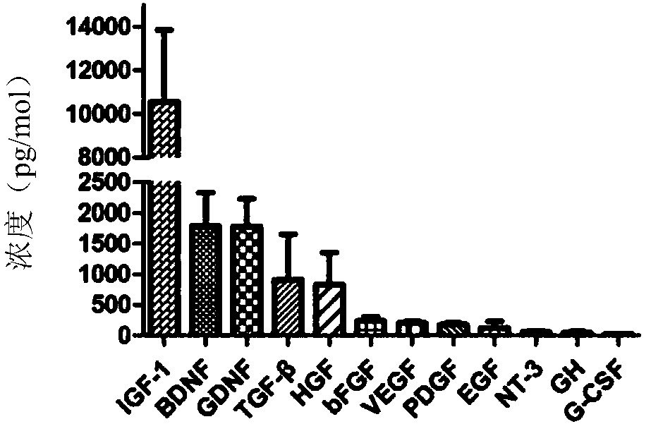
[0042] The obtained fat extract was used to detect the content of growth factors, including IGF-1, BDNF, GDNF, TGF-β, HGF, VEGF, bFGF, PDGF, EGF, NT-3, GH and GM, using ELISA immunosorbent assay kit - Cytokines such as CSF.

[0043] ELISA analysis was carried out on the biomaterials obtained from fats from three donors, and the results showed that IGF-1 (11400±2300pg / mL), BDNF (1860±500pg / mL), GDNF (1820±430pg / mL) in the biomaterials were analyzed by ELISA. ), TGF-β(1020±680pg / mL), HGF(685±384pg / mL), VEGF(229±23pg / mL), bFGF(229±67pg / mL), PDGF(175±30pg / mL) and EGF (139±99pg / mL) is relatively high, and contains trace amounts of NT-3, GH and GM-CSF (see figure 1 ).

Embodiment 3

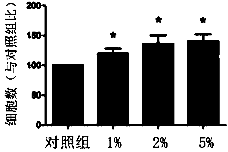
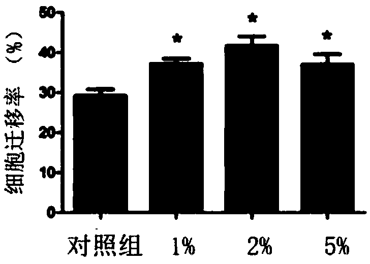
[0044] Example 3 Effects of Biomaterials on the Proliferation of Human Umbilical Vein Endothelial Cells

[0045] Human umbilical vein endothelial cells (Sciencell, U.S.) were added with high-glucose DMEM (Gibco, U.S.) to make a cell suspension, and then inoculated in high-glucose DMEM medium containing 10% fetal bovine serum (HYCLONE, U.S.) , Passed once every 3 days, and the 3rd and 4th passage cells were used in the experiment.

[0046] Human umbilical vein endothelial cells were inoculated on a 96-well plate at a density of 1000 cells per well, and fat extracts of different concentrations (1v / v%, 2v / v% and 5v / v%) were added, each concentration was set at 6 After culturing for 72 hours, CCK8 kit (Beyotime Company, USA) was used to detect cell proliferation and determine the OD value. The experiment was repeated three times to obtain the average value and standard deviation.

[0047] 72 hours after the cells were treated with biological materials, the number of cells detecte...


PUM


Abstract

The invention discloses a biological material, which is taken from adipose tissue and is liquid not containing lipid droplets and cells. The biological material has the effect of promoting angiogenesis and reduces possible side effects and immunogenicity, a preparation method is simple and convenient, complex equipment is not needed, chemical reagents are not involved, safety is high, adding of chemical protection agents is not needed, cryopreservation is easy, and biological activity is maintained.

Description

technical field [0001] The invention relates to a biological material, in particular to an extract separated from adipose tissue, which has the effect of promoting angiogenesis and is suitable for wound repair and the like. Background technique [0002] Angiogenesis plays an important role in the process of regeneration and repair of damaged tissue, especially in ischemic diseases such as myocardial infarction and cerebral infarction. Rapid restoration of blood supply to the injured area is often the decisive factor affecting the prognosis of the disease. In the field of plastic surgery, the survival of free skin flap transplantation, the survival of granular fat grafting, and the healing of skin ulcers caused by diabetes are all closely related to angiogenesis. A large number of studies have shown that the treatment of promoting angiogenesis can significantly improve tissue survival and wound repair. Effect. Traditional local injection of pro-angiogenic growth factors has ...

Claims


Application Information

Patent Timeline
IPC IPC(8): A61L15/40A61L15/42A61L27/36A61L27/50A61P9/10
CPCA61L15/40A61L15/42A61L27/3604A61L27/3641A61L27/3683A61L27/3679A61L27/50A61L27/507A61K47/44A61P9/10A61L2430/40A61L2430/22A61L2300/412A61L2300/414A61L2300/30
Inventor 李伟张文杰于子优蔡宜佐郑红洁邓明武王祥盛徐宇达
Owner 上海金致投资管理有限公司
Why Eureka
Try Eureka