SERS Detection Method Based on Hydroxyapatite Nanoparticle Adsorbed Protein

A technology of hydroxyapatite and nanoparticles, which is applied in the field of protein analysis and detection, can solve the problems of large sample volume, expensive consumables, and complicated steps, and achieve the effects of less total protein, low experimental cost, and simple operation

Active Publication Date: 2022-04-12
FUJIAN NORMAL UNIV
View PDF8 Cites 0 Cited by
  • Summary
  • Abstract
  • Description
  • Claims
  • Application Information

AI Technical Summary

Problems solved by technology

The transfer efficiency of this method is low, usually only a small part of the protein (10%-20%) in the gel can be transferred, and the operation steps are cumbersome
The above method requires a large amount of samples, expensive consumables, complicated steps, long time-consuming, and expensive, so it is difficult to widely promote

Method used


Image

  • SERS Detection Method Based on Hydroxyapatite Nanoparticle Adsorbed Protein
  • SERS Detection Method Based on Hydroxyapatite Nanoparticle Adsorbed Protein
  • SERS Detection Method Based on Hydroxyapatite Nanoparticle Adsorbed Protein

Examples


Embodiment 1

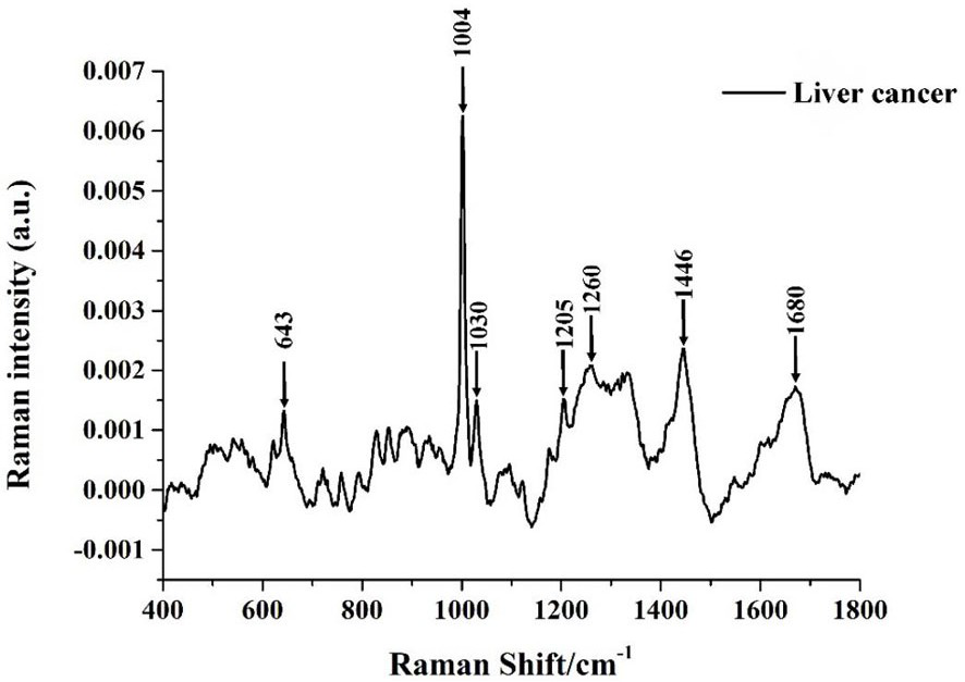
[0038] Surface-enhanced Raman spectroscopy detection of HAP5-adsorbed liver cancer serum protein based on silver nanoparticles substrate

[0039] Weigh 10 mg of HAP5 powder and add it to a 1.5 mL polypropylene centrifuge tube, add 1 mL of distilled water to the centrifuge tube, and ultrasonically disperse it for 10 minutes. Mix evenly, and then place the mixed solution at 100 rpm and incubate for 24 hours at 37° C. in a table-top shaking incubator. After 24 hours, centrifuge the incubated solution at 8000rpm for 10min and discard the supernatant. Add 100µL of acid diluted 10 times to the centrifuge tube that retains the precipitate, adjust the pH value to 1.5, shake it well, let it stand for 5min, and then set it at 6000rpm. After 30s of centrifugation, 10 µL of the supernatant was extracted, and the extracted supernatant and 10 µL of silver nanoparticles were fully stirred, placed on an aluminum sheet with a purity of 99.99%, and dried at room temperature for SERS spectrum de...

Embodiment 2

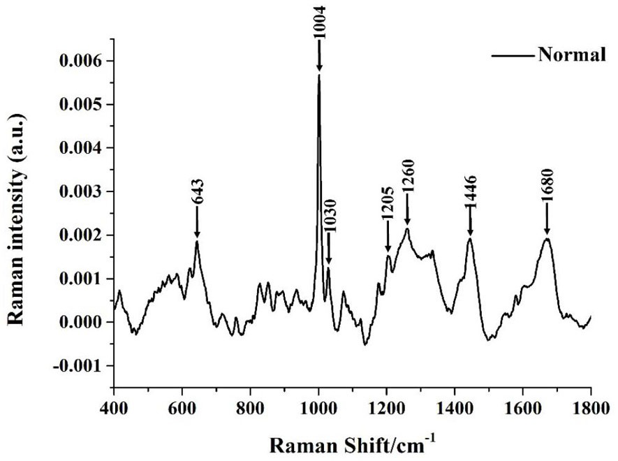
[0041] Surface-enhanced Raman Spectroscopy Detection of HAP5 Adsorbed Normal Human Serum Protein Based on Gold Nanoparticle Substrate

[0042] The method for HAP5 to adsorb normal human serum and release protein is the same as in Example 1. Take 5 µL of gold nanoparticles and 5 µL of the final supernatant, mix them, place them on an aluminum sheet with a purity of 99.99%, dry them at room temperature, and perform SERS Spectral detection. The excitation wavelength of the laser used for detection is 785nm, and the spectral range is 400-1800cm -1 , the power is 10mW, the integration time is 30s, and the integration times are 2 times, the SERS spectrum of normal human serum protein is detected as figure 2 shown.

Embodiment 3

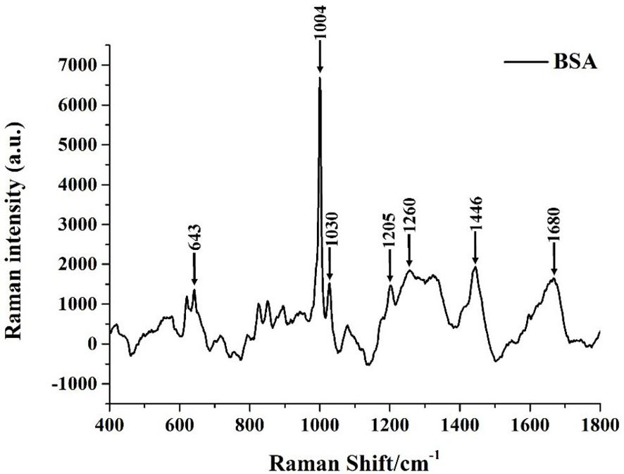
[0044] Surface-enhanced Raman Spectroscopy Detection of HAP5 Adsorbed BSA Based on Silver Nanoparticles Substrate

[0045] Weigh 0.1g of BSA and dissolve it in a certain amount of distilled water. After it dissolves naturally, dilute it in a 50mL volumetric flask to obtain a 1mg / mL BSA protein solution. Weigh 20mg of HAP5 powder and add it to a 1.5mL polypropylene centrifuge tube, add 2mL distilled water to the centrifuge tube, ultrasonically disperse for 10min, measure 1mg / mL BSA solution 1mL, add it to the centrifuge tube after ultrasonic dispersion and shake fully until Mix evenly, and then place the mixed solution at 100 rpm and incubate for 24 hours at 37° C. in a table-top shaking incubator. The method for HAP5 to adsorb BSA and release protein is the same as that in Examples 1 and 2. Take 20 µL of silver nanoparticles and 20 µL of the final extracted supernatant, mix them, put them on a 99.99% pure aluminum sheet, dry them at room temperature, and perform SERS spectrum...


PUM

PropertyMeasurementUnit
wavelengthaaaaaaaaaa
particle diameteraaaaaaaaaa

Abstract

The invention discloses a surface-enhanced Raman spectrum detection method based on hydroxyapatite nanoparticle adsorption protein, in which hydroxyapatite nanoparticle and biological sample are mixed and incubated to absorb protein in the sample, and acid is added to adjust the pH value to make the protein The surface of the hydroxyapatite nanoparticles falls off, and the supernatant containing the exfoliated protein is extracted and mixed with the SERS nano-active material, and the surface-enhanced Raman spectrum of the protein of the biological sample is detected by using the SERS technology, and the surface-enhanced Raman spectrum of the protein of the biological sample is established. Database; through the post-processing and analysis of the data, the characteristic peaks of the surface-enhanced Raman spectrum corresponding to different biological sample proteins are obtained. The invention has the characteristics of rapidity, simple operation, low cost and the like, and can effectively detect the surface-enhanced Raman spectrum of proteins in different biological samples.

Description

technical field [0001] The invention belongs to the field of protein analysis and detection, and in particular relates to a surface-enhanced Raman spectrum detection method based on hydroxyapatite nanoparticle adsorption protein. Background technique [0002] Raman spectroscopy is a molecular spectroscopy technology developed based on the Raman scattering effect. The frequency difference between the scattered light and the excitation light is called the Raman shift. The Raman shift is only related to the rotation and vibration level of the molecule. . Therefore, Raman spectroscopy can provide chemical fingerprint information of the composition and conformation of biological macromolecules such as proteins, nucleotides and lipids, and then the molecular chemical bond structure and vibration information of the substance can be reflected through the peak assignment, which can be used to analyze the molecular structure This method has the advantages of rapid detection, no damag...

Claims


Application Information

Patent Timeline
Patent Type & Authority Patents(China)
IPC IPC(8): G01N21/65
CPCG01N21/658
Inventor 林居强林雅敏俞允高思琪郑萌萌
Owner FUJIAN NORMAL UNIV
Why Eureka
Try Eureka