Visible-light and near-infrared fluorescent 3D co-imaging endoscope system based on single detector

A single detector and imaging system technology, applied in the fields of endoscopy, medical science, diagnosis, etc., can solve the problems of lack of depth information, operation fluency, increase operation time, etc., to ensure the operation process and improve the success rate of operation , small size effect

Inactive Publication Date: 2020-02-28
CHANGCHUN INST OF OPTICS FINE MECHANICS & PHYSICS CHINESE ACAD OF SCI
View PDF7 Cites 7 Cited by
  • Summary
  • Abstract
  • Description
  • Claims
  • Application Information

AI Technical Summary

Problems solved by technology

This working mode has brought troubles to the fluency of the operation and increased the operati

Method used


Image

  • Visible-light and near-infrared fluorescent 3D co-imaging endoscope system based on single detector
  • Visible-light and near-infrared fluorescent 3D co-imaging endoscope system based on single detector
  • Visible-light and near-infrared fluorescent 3D co-imaging endoscope system based on single detector

Examples


Embodiment Construction

[0031] The technical solution of the present invention will be further described below in conjunction with the accompanying drawings.

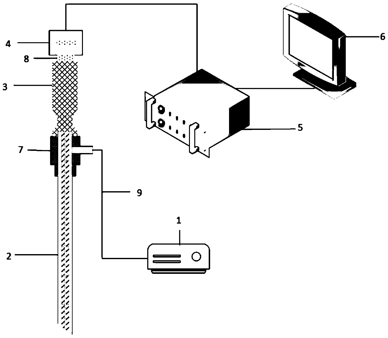
[0032] Such as figure 1 As shown, the visible light and near-infrared fluorescence 3D co-imaging endoscope system based on a single detector of the present invention includes a visible light near-infrared excitation light source 1, a binocular endoscopic imaging system 2, an optical relay image transfer system 3, and an image sensor Module 4 , image processing fusion module 5 , 3D image display system 6 , first fixing part 7 , connecting part 8 and first optical fiber 9 . The binocular endoscopic imaging system 2, the optical relay image transfer system 3 and the image sensor module 4 are the main parts of the endoscopic system, the visible light and near-infrared excitation light source 1, the image processing fusion module 5 and the 3D image display system 6 are the endoscopic Peripherals of the mirror system.

[0033] In the above-mention...


PUM

PropertyMeasurementUnit
Diameteraaaaaaaaaa
Center thicknessaaaaaaaaaa
Diameteraaaaaaaaaa

Abstract

The invention discloses a visible-light and near-infrared fluorescent 3D co-imaging endoscope system based on a single detector, and belongs to the technical field of endoscope imaging systems, achieving simultaneous acquisition and display of visible-light and near-infrared fluorescent 3D images. The 3D co-imaging endoscope system comprises a visible-light near-infrared excitation light source, abinocular endoscope imaging system, an optical relay inversion system, an image sensor module, an image processing fusion module and a 3D image display system. The endoscope system has the advantagesthat two images with horizontal parallax are received by the RGB-NIR detector to achieve acquisition of the 3D images, so that the visible-light colored 3D image and the near-infrared fluorescent 3Dimage are acquired at the same time; the system structure is simple, and the size is small; operation interruption for status switching is not needed, and smooth operation processes are ensured; the position and size of lesion tissue can be sensed by doctors directly, and accordingly, the success rate of operations is greatly increased.

Description

technical field [0001] The invention belongs to the technical field of endoscope imaging systems, in particular to a single-detector-based visible light and near-infrared fluorescence 3D co-imaging endoscope system. Background technique [0002] Endoscopic technology is a comprehensive technology that integrates advanced disciplines such as optics, precision manufacturing, image processing, photoelectric information, materials and bioengineering. The emergence of endoscope is an important milestone in the history of medical technology development. It makes minimally invasive surgical examination and operation possible, greatly reduces the harm to patients and effectively controls the risk of surgery. Since the appearance of endoscope, it has experienced the development of rigid tube endoscope, half-curved endoscope, fiber endoscope and electronic endoscope. In recent years, endoscopes combining high-definition video technology and 3D video technology have been proposed one ...

Claims


Application Information

Patent Timeline
IPC IPC(8): A61B1/06A61B1/04
CPCA61B1/00163A61B1/04A61B1/043A61B1/06
Inventor 史成勇张红鑫王泰升
Owner CHANGCHUN INST OF OPTICS FINE MECHANICS & PHYSICS CHINESE ACAD OF SCI
Why Eureka
Try Eureka