
Trichomonas vaginalis fluorescence immunochromatographic assay kit and preparation method thereof

A technique of fluorescence immunochromatography and Trichomonas vaginalis, applied in measuring devices, analysis materials, instruments, etc., can solve the problems of large system error, unfavorable CV control, complicated preparation process, etc., and achieve uniform samples, sophisticated equipment, high The effect of detection sensitivity

Inactive Publication Date: 2020-04-17
江苏美克医学技术有限公司
View PDF18 Cites 0 Cited by
  • Summary
  • Abstract
  • Description
  • Claims
  • Application Information

AI Technical Summary

Problems solved by technology

[0004] Chinese patent CN107132353A discloses a detection kit for group B streptococci and its preparation method. Although it can also give quantitative detection results, it uses the ABS system, the preparation process is complicated, and two strains of antibodies are required to be labeled separately. There are two Combined pads, two sample processing solutions, need to be mixed in proportion when used, the system error is large, which is not conducive to the control of CV

Method used


Image

  • Trichomonas vaginalis fluorescence immunochromatographic assay kit and preparation method thereof
  • Trichomonas vaginalis fluorescence immunochromatographic assay kit and preparation method thereof
  • Trichomonas vaginalis fluorescence immunochromatographic assay kit and preparation method thereof

Examples


Embodiment 1

[0050] Embodiment 1: Conjugate pad preparation

[0051] 1. Buffer replacement: take 1mg of 200nm fluorescent microspheres, add 1mL of 100mM MES, pH5.5-6.0 (the following MES are all 100mM), centrifuge (14000g for 30min). Add 500 μL MES to resuspend by ultrasonication and set aside.

[0052] 2. Activation: Add 0.06 mg NHS, mix well, add 0.1 mg EDC, add 500 μL MES, and rotate at 37°C for 30 min to 60 min (the speed of the mixing rotator is set to 30-50).

[0053] 3. Cleaning: After activation, add 1mL MES, centrifuge (14000g for 30min), remove the supernatant, add 500μL MES for ultrasonic resuspension, repeat once, add 50mM HEPES, pH8.0 (the following HEPES are all 50mM, pH8.0) 500μL , ultrasonically resuspend, add 1 mL HEPES for centrifugation, add 500 μL HEPES ultrasonically resuspend and set aside.

[0054] 4. Antibody reaction: add 0.03mg of antibody (antibody HEPES pre-dialyzed), mix well and react at 37°C for 1-4hr.

[0055] 5. Blocking 1: Add 7 μL of 2M Gly and 14 μL o...

Embodiment 2

[0058] Embodiment 2: Conjugate pad preparation

[0059] The added amount of the antibody is 0.05 mg, the added amount of the rabbit IgG is 1 mg, and the rest of the steps are the same as in Example 1.

Embodiment 3

[0060] Embodiment 3: Conjugate pad preparation

[0061] The added amount of the rabbit IgG is 0.05 mg, and the added amount of the rabbit IgG is 2 mg. The rest of the steps are the same as in Example 1.


PUM


Abstract

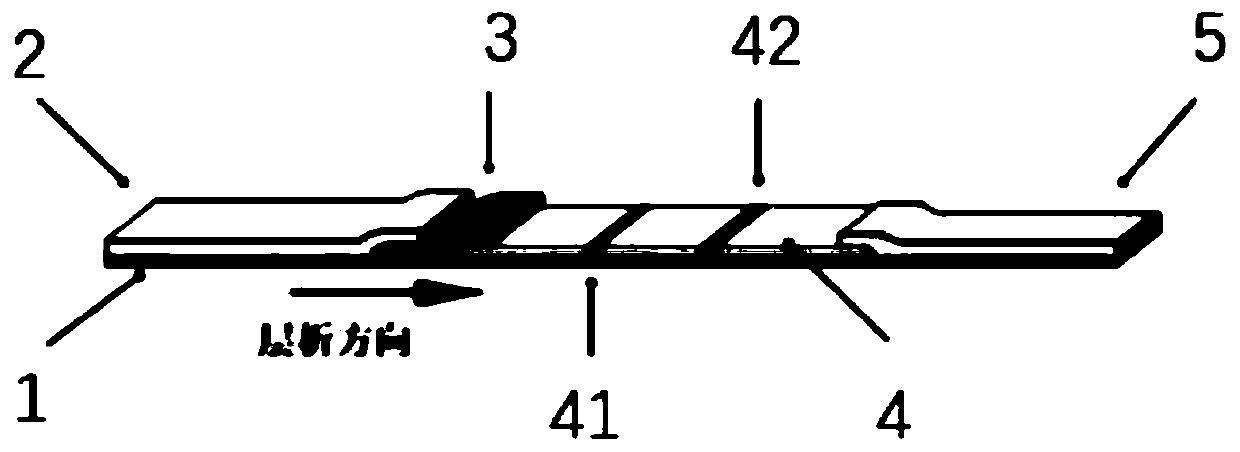
The invention provides a trichomonas vaginalis fluorescence immunochromatographic assay kit and a preparation method thereof. The trichomonas vaginalis fluorescence immunochromatographic assay kit comprises a detection card, an SD card and a sample diluent, wherein the detection card is composed of a shell and an immunochromatographic test strip, the immunochromatographic test strip is positionedin the shell and comprises a bottom plate, a sample pad, a combination pad, a nitrocellulose membrane and a sample suction pad are sequentially arranged on the bottom plate along the length direction,an anti-trichomonas vaginalis monoclonal antibody marked by fluorescent microspheres is fixed on the combination pad, a detection T line and a quality control C line are arranged on the nitrocellulose membrane, the T line is coated with a monoclonal antibody paired with the monoclonal antibody marked by the fluorescent microspheres, and the C line is coated with a secondary antibody paired with the monoclonal antibody marked by the fluorescent microspheres. The kit disclosed by the invention is higher in detection sensitivity, accuracy and precision, and has important significance for dynamically monitoring the illness state of a patient, performing prognosis evaluation, detecting recurrence and guiding clinicians to make important medical decisions in time.

Description

technical field [0001] The invention belongs to the technical field of microbial immune detection, and more specifically relates to a fluorescent immunochromatographic assay kit for Trichomonas vaginalis and a preparation method thereof. Background technique [0002] Trichomonas vaginalis is a flagellate parasitic in the human vagina and urinary tract, mainly causing trichomonal vaginitis and urethritis, and is a sexually transmitted infectious disease. [0003] Trichomonas vaginalis is a parasite, but it is invisible to the naked eye. This Trichomonas is broadly pear-shaped or oval, with a length of 10-30 μm and a width of 10-20 μm. There are 4 roots on the head as long as the body. Flagella, this trichomonad can be clearly seen under a microscope. Trichomonas has strong adaptability to different environments, it can grow and reproduce at 25°C-42°C, it can survive for 21 days at a low temperature of 3°C-5°C, and it can still survive for 20-60 minutes at 46°C. It can also ...

Claims


Application Information

Patent Timeline
IPC IPC(8): G01N33/577G01N33/569G01N33/558G01N33/533
CPCG01N33/577G01N33/56905G01N33/558G01N33/533
Inventor 卢铖
Owner 江苏美克医学技术有限公司
Learn More