A radiomics-based imaging tool to monitor tumor-lymphocyte infiltration and outcome in cancer patients treated by anti-PD-1/PD-L1

A radiomics and cellular technology, applied in the field of biomarkers, can solve the problem of low level of evidence

Pending Publication Date: 2020-05-01
INSTITUT GUSTAVE ROUSSY
View PDF8 Cites 13 Cited by
  • Summary
  • Abstract
  • Description
  • Claims
  • Application Information

AI Technical Summary

Problems solved by technology

[0008] Four published studies evaluated imaging as a biomarker of immune infiltration or immune pathways [54, 59-61], but no patients were treated with immunotherapy and only one validated its radiomics signature in an independent cohort [ 60]
However, the level of evidence from these studies was low, with only one study having an external validation cohort

Method used


Image

  • A radiomics-based imaging tool to monitor tumor-lymphocyte infiltration and outcome in cancer patients treated by anti-PD-1/PD-L1
  • A radiomics-based imaging tool to monitor tumor-lymphocyte infiltration and outcome in cancer patients treated by anti-PD-1/PD-L1
  • A radiomics-based imaging tool to monitor tumor-lymphocyte infiltration and outcome in cancer patients treated by anti-PD-1/PD-L1

Examples


Embodiment 1

[0164] Example 1: Contrast-Enhanced Computed Tomography (CT) Imaging of Head and Neck Cancer

[0165] training group

[0166] Clinical and imaging data from the head and neck squamous cell carcinoma cohort provided by The Cancer Imaging Archive (http: / / www.cancerimagingarchive.net / ) (TCIA-HNSC) were used [35, 36]. Baseline contrast-enhanced computed tomography (CT) imaging with slice thickness equal to or less than 5 mm and acquired using a 120KVP was reviewed. After excluding patients with only postoperative imaging or artifacts at the tumor level, no gantry-tilt (for technical compatibility with our texture imaging software), and no available genomic data, 57 patients were included. patient.

[0167] The corresponding genomic data were obtained from the Cancer Genome Altas (TCGA) Portal (https: / / portal.gdc.cancer.gov / ), which included the use of Illumina HiSeq RNASeqV2 (Illumina, San Diego, California, USA ) and quantified 20,530 genes using the RPKM (Reads Per Kilobase M...

Embodiment 2

[0202] Example 2: Contrast-enhanced CT-based radiomics labeling of different solid tumors

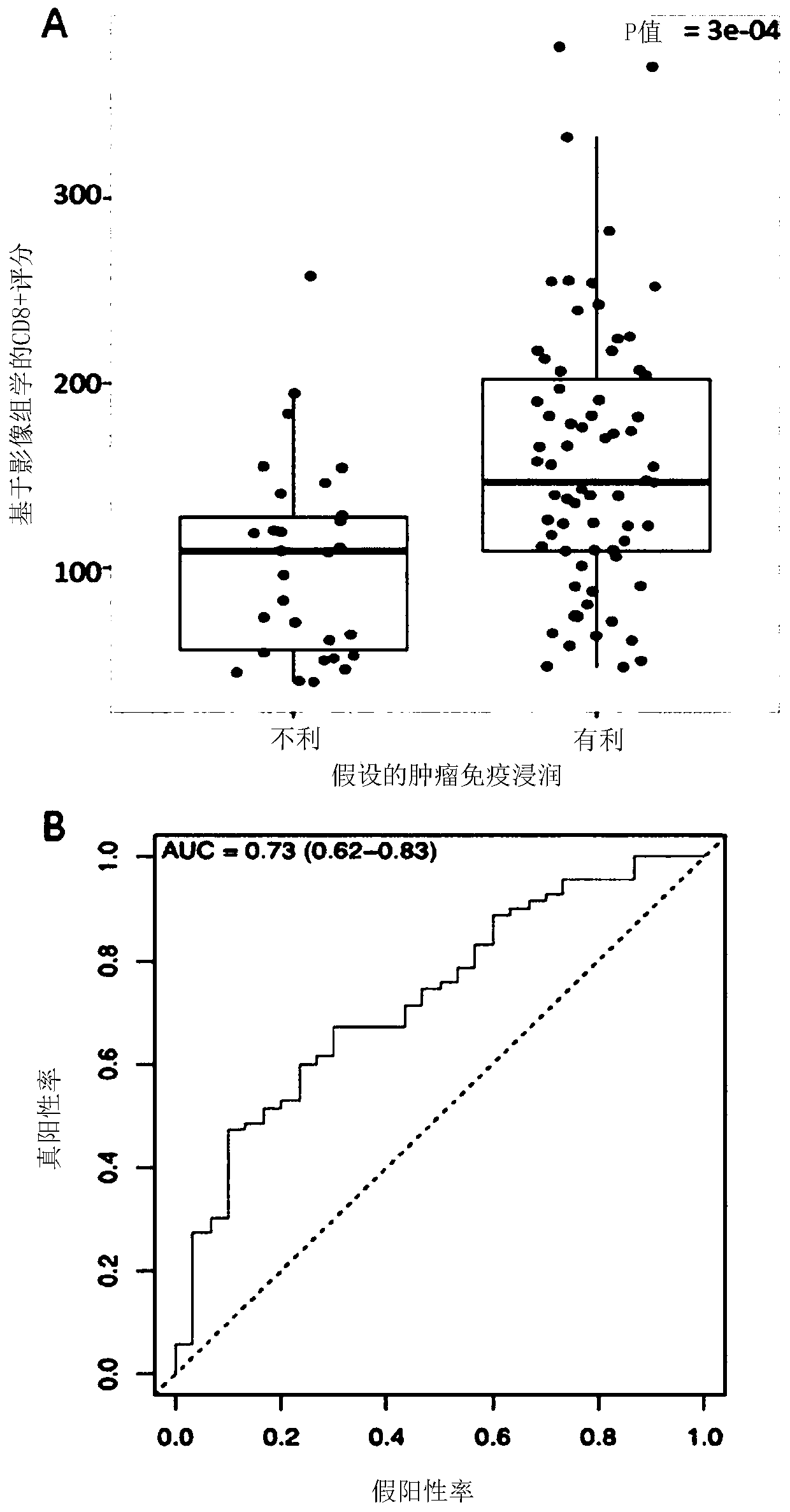
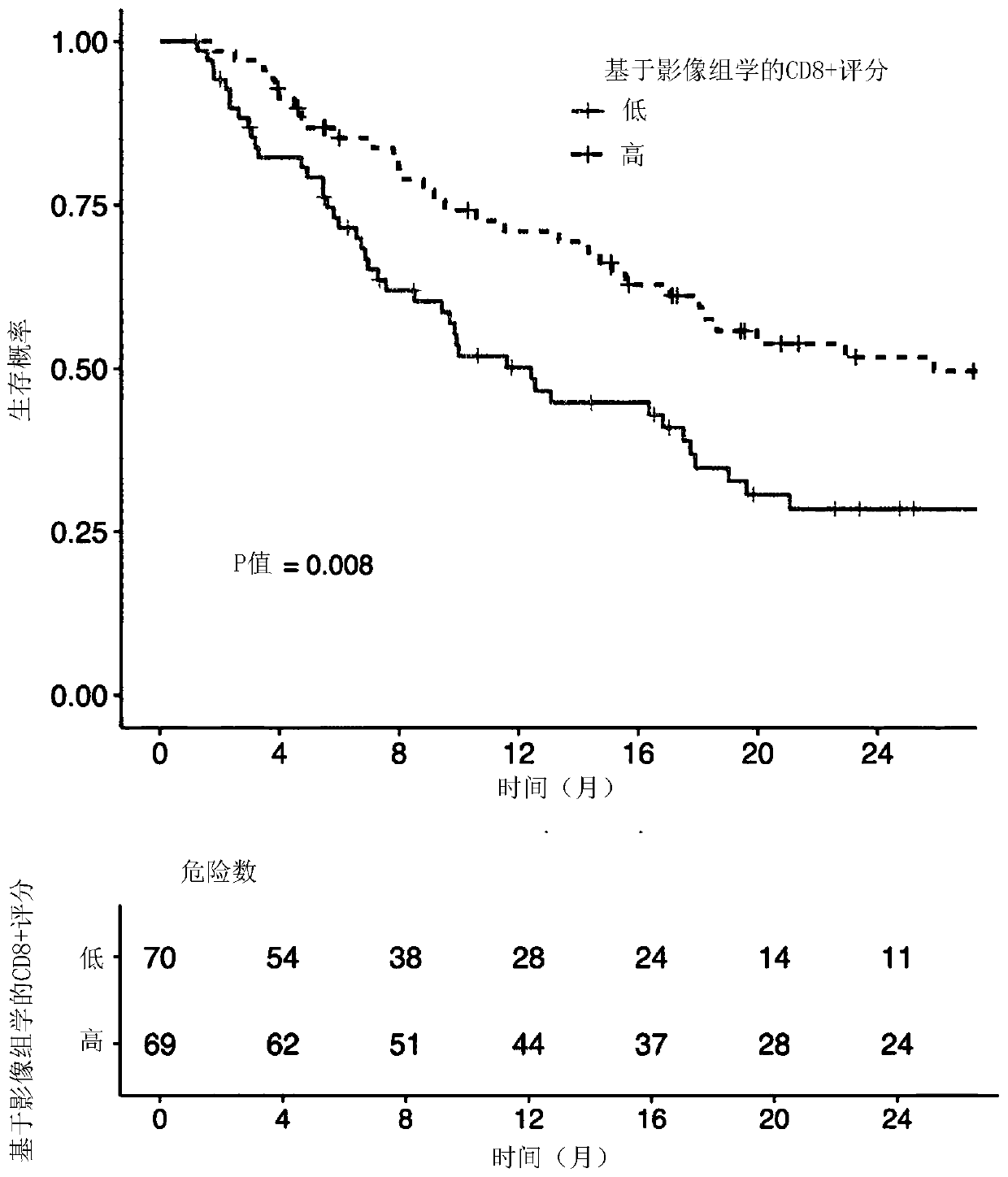
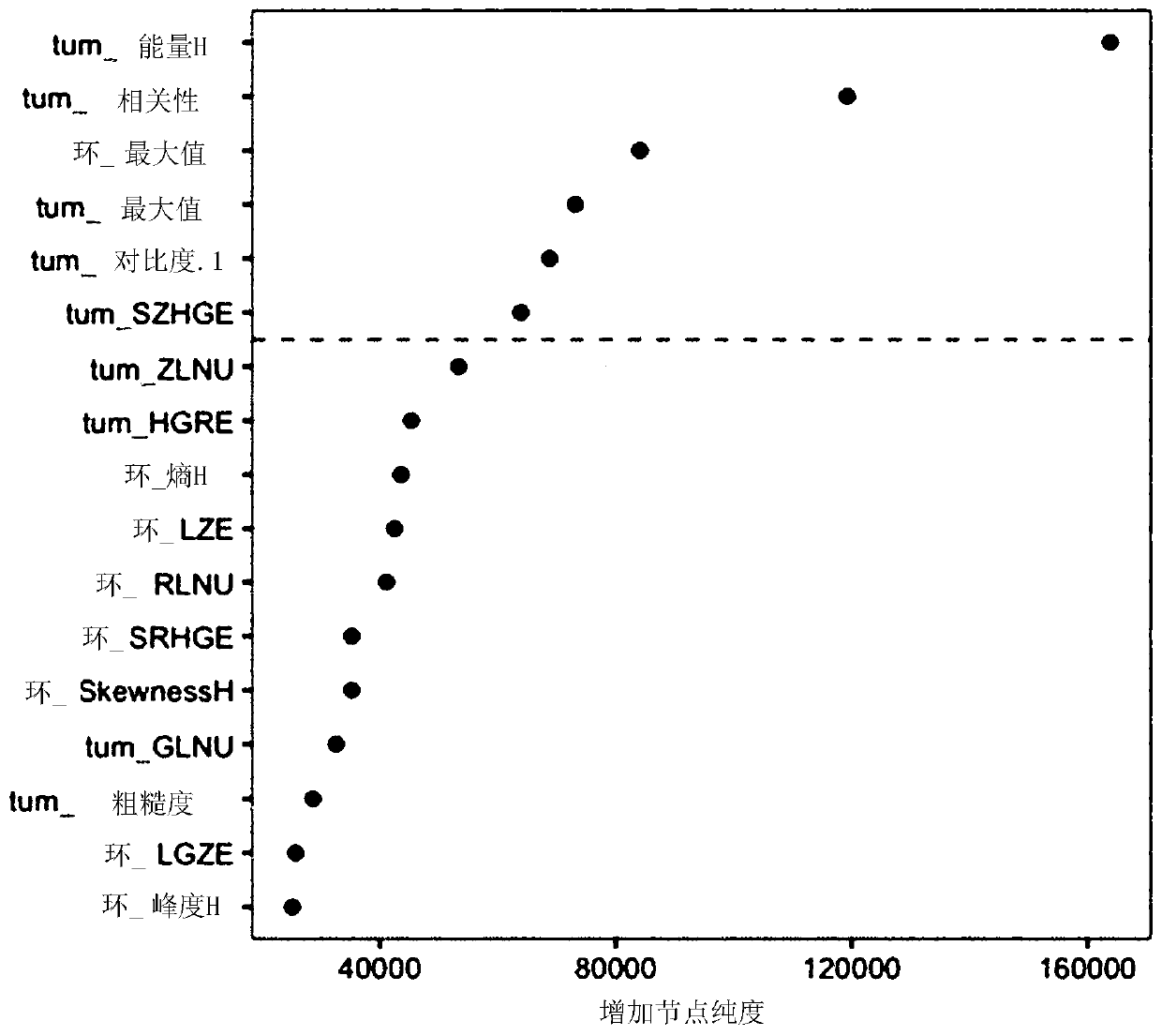
[0203] Radiomics features were extracted from contrast-enhanced CT in 135 patients with advanced solid malignancies in the prospective trial MOSCATO. For each patient, RNA-seq data were used to quantify CD8 T cells. From 84 variables (78 radiomics features, 5 location variables, 1 technology variable), radiomics-based predictors of CD8 T cell expression were constructed using Elastic Net. The primary goal was to confirm the relationship of this predictor to gene expression in an independent cohort of 119 patients from The Cancer Genome Atlas (TCGA). Two separate cohorts of patients with solid tumors were used to assess this predictor: 100 patients were assumed to have tumors that were either immune primed (dense CD8 T cell infiltration) or immune desert (limited CD8 T cell infiltration) , to analyze the correlation with the immune phenotype, and 137 patients treated in a phase 1 trial...


PUM

PropertyMeasurementUnit
thicknessaaaaaaaaaa

Abstract

The present invention proposes a radiomics-based biomarker for detecting the presence and the density of tumor infiltrating CD8 T-cells in a solid tumor without having to use any biopsy of said tumor.The invention also proposes to use this information to assess the immune phenotype of said solid tumor. In a particular embodiment, the invention proposes to prognose the survival and / or the treatment efficiency of cancer patients treated with immunotherapy such as anti-PD-1 / PD- L1 monotherapy.

Description

technical field [0001] The present invention proposes a radiomics-based biomarker for detecting the presence and density of tumor-infiltrating CD8 T cells in solid tumors without any biopsy of the tumor. The invention also proposes to use this information to assess the immunophenotype of said solid tumor. In a specific embodiment, the present invention proposes to predict survival and / or therapeutic efficacy of cancer patients treated with immunotherapy, such as anti-PD-1 / PD-L1 monotherapy. Background technique [0002] Medical image processing and analysis (also known as radiomics) is a promising and rapidly developing discipline [1]. Classical analysis of medical images such as computed tomography (CT), magnetic resonance imaging (MRI) or PET (positron emission tomography) is based on the visual interpretation of simple features such as tumor size and overall tumor shape, which The new method analyzes images and computationally converts them into quantitative complex dat...

Claims


Application Information

Patent Timeline
Patent Type & Authority Applications(China)
IPC IPC(8): G01N33/50G06T7/00G16H30/40G16H50/20
CPCG01N33/5005G01N2800/52G01N2800/7028G06T7/0012G16H30/40G16H50/20G06T2207/10081G06T2207/10104G06T2207/10088G06T2207/30096G16H30/20G06T2207/10072G01N33/505G01N33/574A61B6/5217
Inventor C·费尔泰E·J·利姆基尼R·孙E·多伊奇
Owner INSTITUT GUSTAVE ROUSSY
Why Eureka
Try Eureka