Supercharge Your Innovation With Domain-Expert AI Agents!

Method for processing human joint data provided based on tomography technology

A technology for human joint and tomographic imaging, which is applied in the field of medical image processing and can solve problems such as affecting doctors' judgment.

Inactive Publication Date: 2020-05-05
ZHONGNAN HOSPITAL OF WUHAN UNIV
View PDF2 Cites 3 Cited by
  • Summary
  • Abstract
  • Description
  • Claims
  • Application Information

AI Technical Summary

Problems solved by technology

Moreover, due to the influence of the patient's posture during the shooting and the doctor's operation of the shooting equipment, there may be some differences in the spatial positions of the sequences obtained by the two MRI scans before and after, and for very thin articular cartilage, even a subtle Differences may also affect physician judgment

Method used


Image

  • Method for processing human joint data provided based on tomography technology
  • Method for processing human joint data provided based on tomography technology
  • Method for processing human joint data provided based on tomography technology

Examples


Embodiment 1

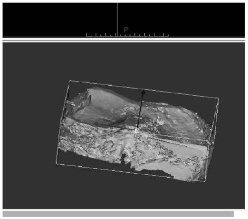
[0079] refer to figure 2 According to one embodiment of the present invention, a method for processing a magnetic resonance scan sequence taken for a human knee joint to obtain data for establishing a three-dimensional articular cartilage model includes:

[0080] Step 1. Use the MRI scanner to collect image data of the knee joint.

[0081] When collecting images of the knee joint, the patient can be prompted to lie down and adjust the body position, so that the degree of flexion and extension of the knee joint, placement, and the rotation angle compared to the scanner are fixed, so that the same image can be taken every time. The same area of ​​the knee joint of the person in the same posture is photographed from the same angle.

[0082] According to an embodiment of the present invention, in order to facilitate the acquisition of accurate articular cartilage data, an image analysis program may be used to perform image processing such as denoising on the obtained scan sequence...

Embodiment 2

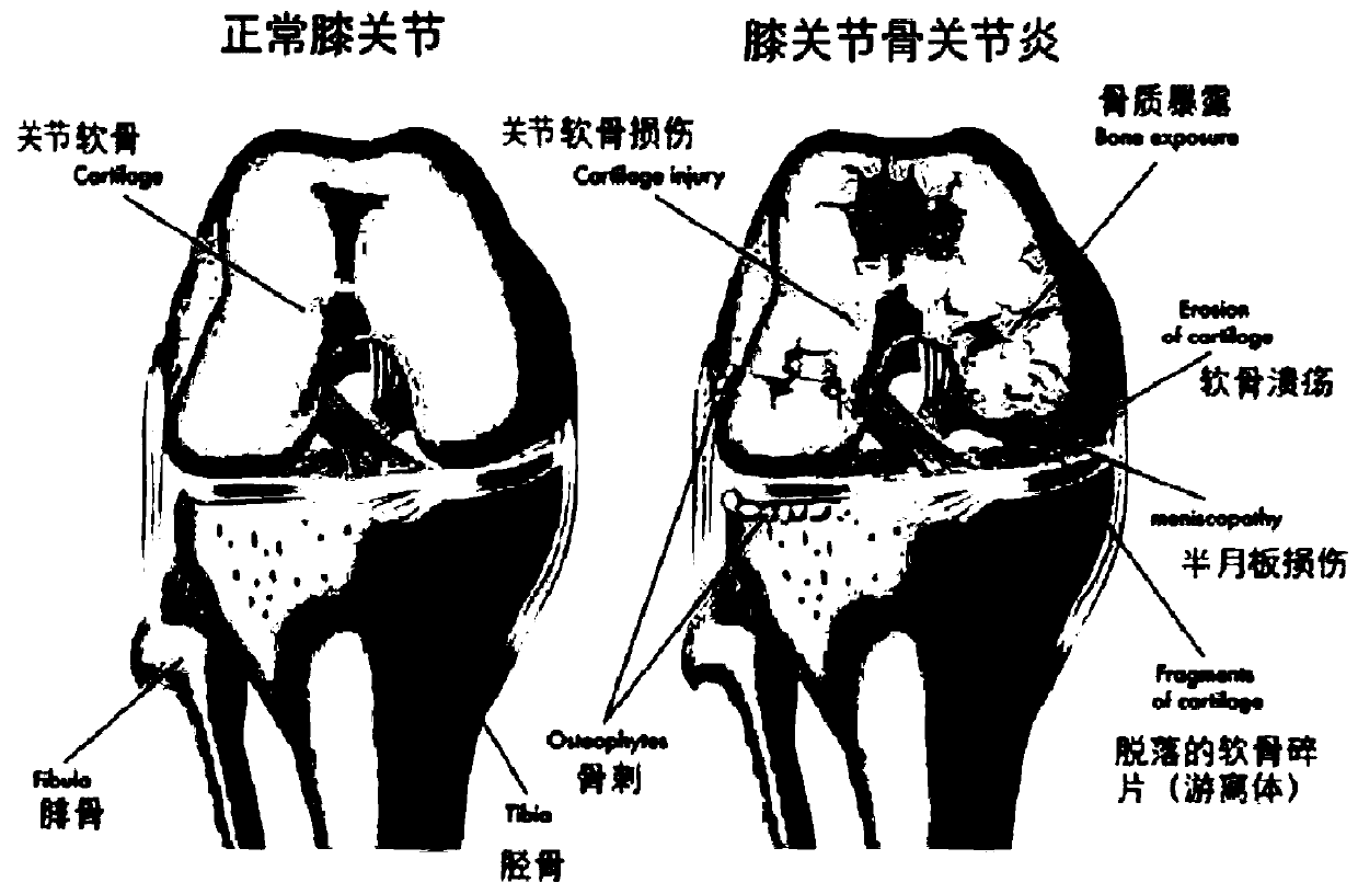
[0107] The inventors found that in the course of medical practice, in order to track the progress of a patient's disease to determine the appropriate treatment, or to study whether the articular cartilage of a patient is repaired for a period of time after the treatment with adipose-derived mesenchymal stem cells , it is necessary to compare the condition of the articular cartilage of the patient in the two periods.

[0108] Figure 3e A schematic diagram of the three-dimensional model of the cartilage attached to the osteogenesis on both sides of the right knee joint obtained by the method of before treatment, 12 weeks after treatment, and 24 weeks after treatment is shown. It can be seen that based on this method, it is convenient to conduct a qualitative analysis of the cartilage wear conditions captured several times. However, in order to quantitatively compare the cartilage wear conditions, it is necessary to spatially analyze the cartilage of the same joint obtained by ...


PUM


Abstract

The invention provides a method for processing human joint data provided based on a tomography technology. The method comprises the following steps: 1) acquiring human body joint data I correspondingto a scanning sequence of first tomography and human body joint data II corresponding to a scanning sequence of second tomography for the same joint part of the same photographed person; and 2) registering the cartilage part in the human body joint data I and the cartilage part in the human body joint data II into the same spatial domain based on the osteogenesis parts in the human body joint dataI and the human body joint data II. According to the method, the corresponding relation between the corresponding detection positions of two times of tomography can be determined, so that the data can be used for qualitatively and quantitatively analyzing and comparing articular cartilage images of the same patient in different periods.

Description

technical field [0001] The invention relates to medical image processing technology, in particular to a method for providing data for qualitative and quantitative analysis of articular cartilage. Background technique [0002] Osteoarthritis (OA) is a degenerative joint disease, also known as degenerative arthritis or bone and joint disease, and is a common disease among middle-aged and elderly people. Figure 1a A comparison of a normal knee joint and a typical knee joint with osteoarthritis is shown. From the figure, it can be seen that the patient's articular cartilage has been damaged so that part of the bone is exposed, which is also accompanied by cartilage ulcers and menisci. Injury, fragments of sloughed cartilage exist as loose bodies in the joint. In this case, due to the body's immune response, various inflammations develop. The degeneration of cartilage tissue is the main cause of pain in patients with osteoarthritis. After the change of cartilage tissue, patient...

Claims


Application Information

Patent Timeline
Patent Type & Authority Applications(China)
IPC IPC(8): G06T7/00G06T7/33G06T7/62G06T11/00
CPCG06T2207/10088G06T7/0012G06T7/33G06T7/62G06T11/003G06T2207/30008
Inventor 郑心校徐海波李涛包益桂肖峰阮兆蔡林
Owner ZHONGNAN HOSPITAL OF WUHAN UNIV
Features
  • R&D
  • Intellectual Property
  • Life Sciences
  • Materials
  • Tech Scout
  • Unparalleled Data Quality
  • Higher Quality Content
  • 60% Fewer Hallucinations
Learn More