Fundus blood vessel three-dimensional reconstruction method and device and electronic equipment

A 3D reconstruction and blood vessel technology, applied in 3D modeling, image data processing, instruments, etc., can solve the problems of slow image speed, low resolution, low efficiency and accuracy of 3D reconstruction of fundus blood vessels, etc. High rate, fast effect

Pending Publication Date: 2020-06-05
SHANGHAI FIRST PEOPLES HOSPITAL
View PDF4 Cites 1 Cited by
  • Summary
  • Abstract
  • Description
  • Claims
  • Application Information

AI Technical Summary

Problems solved by technology

However, commonly used fundus imaging instruments have low resolution and slow image generation speed, and need to use contrast agents to obtain retinal choroidal vascular images in vivo, which will lead to low efficiency and accuracy of 3D reconstruction of fundus vessels

Method used


Image

  • Fundus blood vessel three-dimensional reconstruction method and device and electronic equipment
  • Fundus blood vessel three-dimensional reconstruction method and device and electronic equipment
  • Fundus blood vessel three-dimensional reconstruction method and device and electronic equipment

Examples


Embodiment Construction

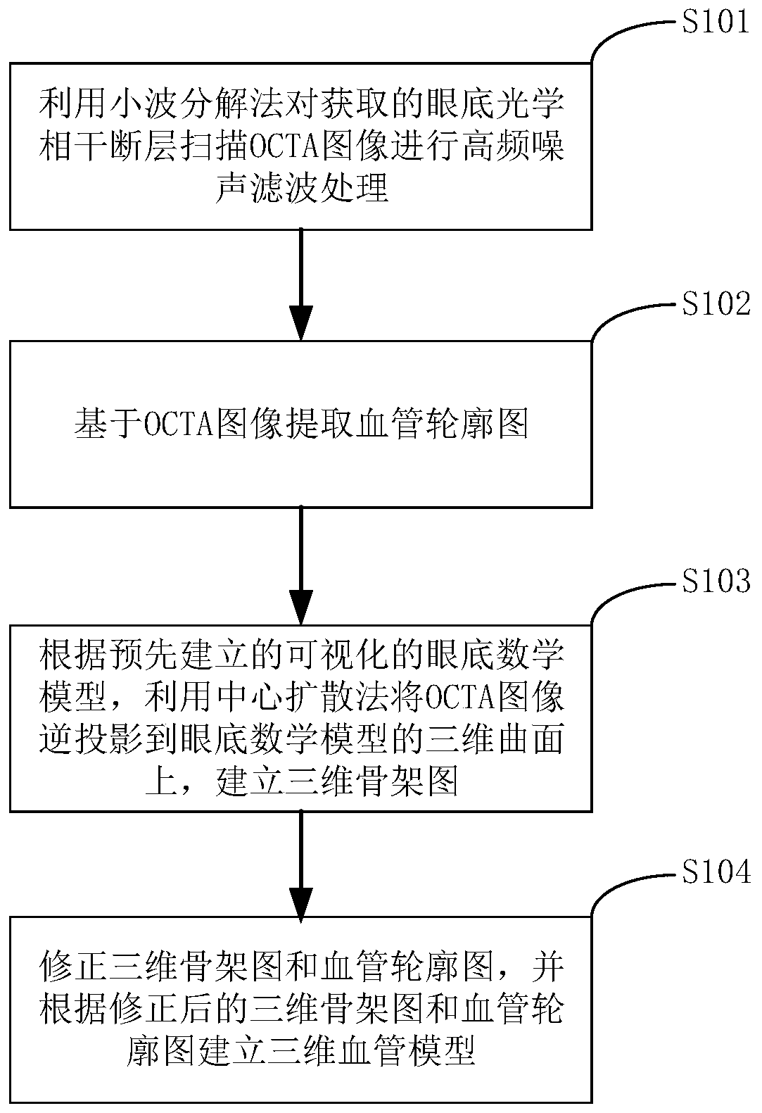
[0036] In order to make the purpose, technical solutions and advantages of the embodiments of the present invention clearer, the technical solutions of the present invention will be clearly and completely described below in conjunction with the accompanying drawings. Obviously, the described embodiments are part of the embodiments of the present invention, not all of them. the embodiment. Based on the embodiments of the present invention, all other embodiments obtained by persons of ordinary skill in the art without making creative efforts belong to the protection scope of the present invention.

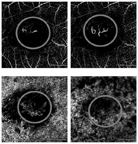
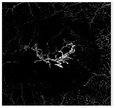
[0037] At present, the commonly used fundus imaging instruments have low resolution and slow image generation speed, and need to use contrast agents to obtain images of retinal choroidal vessels in vivo, which will lead to low efficiency and accuracy of 3D reconstruction of fundus vessels. The Optical Coherence Tomography Angiography (OCTA) technology is a new type of optical non-inv...


PUM


Abstract

The invention provides a fundus blood vessel three-dimensional reconstruction method and device and electronic equipment. The fundus blood vessel three-dimensional reconstruction method comprises thesteps: carrying out high-frequency noise filtering processing on an obtained fundus optical coherence tomography OCTA image through a wavelet decomposition method; extracting a blood vessel contour diagram based on the OCTA image; according to a pre-established visual fundus mathematic model, reversely projecting the OCTA image onto a three-dimensional curved surface of the fundus mathematic modelby utilizing a central diffusion method, and establishing a three-dimensional skeleton diagram; and correcting the three-dimensional skeleton diagram and the blood vessel contour diagram, and establishing a three-dimensional blood vessel model according to the corrected three-dimensional skeleton diagram and blood vessel contour diagram. According to the invention, the efficiency and accuracy offundus blood vessel three-dimensional reconstruction can be improved.

Description

technical field [0001] The invention relates to the technical field of digital image processing, in particular to a method, device and electronic equipment for three-dimensional reconstruction of fundus blood vessels. Background technique [0002] Fundus retinal blood vessels are part of the vascular circulation system of the human body. Many cardiovascular diseases can be diagnosed by observing the changes of retinal blood vessels in the fundus. In the existing methods, a two-dimensional image of the fundus is usually obtained by a fundus imaging device, and a three-dimensional structure schematic diagram of the fundus is drawn using computer technology to reconstruct a three-dimensional model of fundus blood vessels. However, commonly used fundus imaging instruments have low resolution and slow image generation speed, and need to use contrast agents to obtain retinal choroidal vascular images in vivo, which will lead to low efficiency and accuracy of 3D reconstruction of f...

Claims


Application Information

Patent Timeline
Patent Type & Authority Applications(China)
IPC IPC(8): G06T17/00G16H30/40G06T7/00
CPCG06T17/00G16H30/40G06T7/0012G06T2207/30041G06T2207/30101G06T2207/10101
Inventor 孙梦莎汪枫桦宋珉璐孙晓东
Owner SHANGHAI FIRST PEOPLES HOSPITAL
Why Eureka
Try Eureka