Fluorescent probe and preparation method thereof
A fluorescent probe and compound technology, applied in the field of biomedicine, can solve problems such as the impact of diagnostic accuracy, and achieve accurate diagnosis, good tissue penetration, biocompatibility and biodegradability.
- Summary
- Abstract
- Description
- Claims
- Application Information
AI Technical Summary
Problems solved by technology
Method used
Image
Examples
Embodiment 1
[0042] Present embodiment is the preparation of formula (I) compound (PROP, compound 6):
[0043]
[0044] (1) Accurately weigh 0.143 g of compound 1 and 0.214 g of compound 2 and mix, add 10 ml of anhydrous acetonitrile as a solvent, and react at 70° C. for 24 h to obtain compound 3 (compound of formula II).
[0045](2) Weigh 0.181g of compound 4 and 0.202g of 1,3-dibromobutane, add 10ml of anhydrous DMF as a reaction solvent after mixing, and use 200ul of triethylamine as a catalyst, react at room temperature for 12h to obtain compound 5 (formula III compounds).
[0046] (3) Mix 0.303 g of compound 5 and 0.358 g of compound 3, add 200 ul of triethylamine as a solvent, and stir at room temperature for 24 hours to obtain compound 6 (compound of formula I).
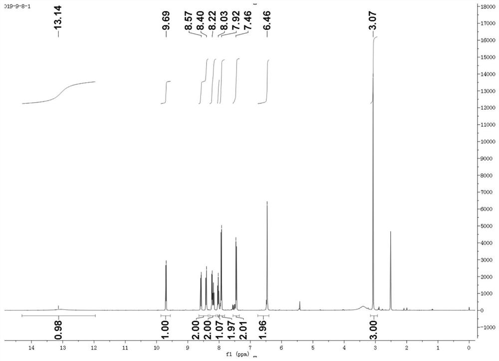
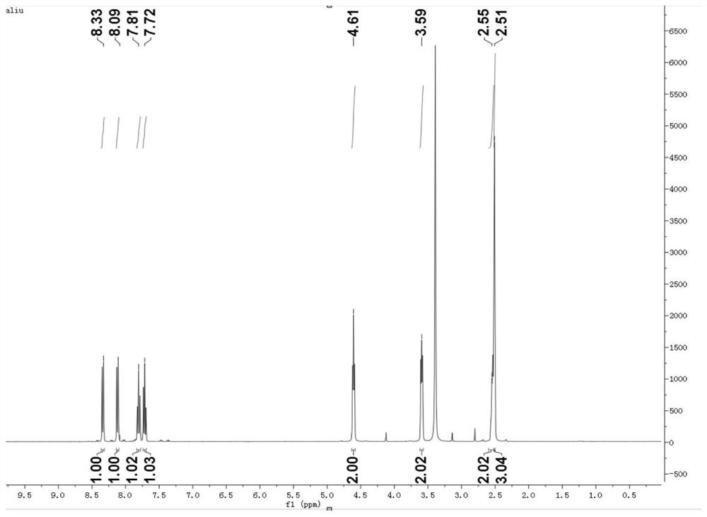
[0047] Depend on Figure 1~4 According to the H NMR spectrum and NMR mass spectrometry results, Compound 3, Compound 5, and Compound 6 were successfully prepared in this example.
Embodiment 2
[0049] This embodiment is the synthesis of fluorescent probe (TPP)
[0050] 1. Through the EDC / NHS technology, 3 mg of transferrin and 0.9 mg of small molecule PROP were coupled at 4°C for 48 hours, and then dialyzed with a dialysis bag for 36 hours to remove free small molecules and collected in the dialysis bag The sample is the TPP obtained from the reaction, which is stored in a 4°C refrigerator for later use, and the above experiments must be completed under dark conditions.
[0051] 2. Prepare 1mg / ml PROP mother solution, and dilute it into 15 different concentrations, the minimum concentration is 0mg / ml, then measure its absorption spectrum in the range of 600-900nm with a UV spectrophotometer, and make a standard curve according to the absorption spectrum ; Measure the absorption spectra of different dosage ratio samples, calculate the PROP concentration in the sample by the standard curve, and then according to the drug loading=(the quality of PROP in the nanoparticle...
Embodiment 3
[0053] This embodiment is TPP toxicity detection
[0054] Select the U87 cells in the logarithmic growth phase, digest and centrifuge, discard the supernatant and resuspend the cells, collect the single cell suspension, perform cell counting, calculate and dilute to an appropriate concentration with complete medium, inoculate in a 96-well plate, and 37°C, 5% CO 2 After culturing for 24 hours under the condition of 95% relative humidity, discard the medium, wash with PBS three times, add different concentrations of free PROP and TPP nanoparticles for cell culture, after incubation for 4 hours, discard the medium, add CCK-8 The fresh medium of the reagent was incubated for 4 hours, the absorption of the sample at 450 nm was detected with a multi-functional microplate reader, and the cell survival rate was calculated by the MTT method.
[0055] From Figure 5 It can be seen that the PROP and TPP small molecule probes have low toxicity and basically no damage to cells, which mee...
PUM
| Property | Measurement | Unit |
|---|---|---|
| wavelength | aaaaa | aaaaa |
Abstract
Description
Claims
Application Information
 - R&D
- Intellectual Property
- Life Sciences
- Materials
- Tech Scout
- Unparalleled Data Quality
- Higher Quality Content
- 60% Fewer Hallucinations
Browse by: Latest US Patents, China's latest patents, Technical Efficacy Thesaurus, Application Domain, Technology Topic, Popular Technical Reports.
© 2025 PatSnap. All rights reserved.Legal|Privacy policy|Modern Slavery Act Transparency Statement|Sitemap|About US| Contact US: help@patsnap.com


