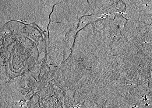
Protein immunogold labeling method for cryoelectron microscope

A cryo-electron microscope, protein technology, applied in the direction of material analysis, instruments, measurement devices, etc. using wave/particle radiation, which can solve problems such as inability to label proteins

Inactive Publication Date: 2020-11-13
XIANGYA HOSPITAL CENT SOUTH UNIV
View PDF9 Cites 2 Cited by
  • Summary
  • Abstract
  • Description
  • Claims
  • Application Information

AI Technical Summary

Problems solved by technology

However, the disadvantage of cryo-electron microscopy is that it can only display the three-dimensional structure of the protein and cannot label the protein.

Method used


Image

  • Protein immunogold labeling method for cryoelectron microscope

Examples


Embodiment 1

[0033] An immunogold labeling protein method for cryo-electron microscopy, the target protein is the thylakoid membrane protein of brown algae, and the specific labeling process is as follows:

[0034] 1) Pretreatment of the grid

[0035] The grid of the cryo-electron microscope is a 200-mesh copper mesh covered with a continuous carbon film. First, the grid is cleaned with pure acetone, and then the grid is treated with a glow discharge instrument, and then the mass fraction is 0.01% poly-L- Cover the grid with lysine solution for 2 hours, remove the poly-L-lysine solution, then soak the grid with 1x PBS for 2 hours, and finally clamp the grid with tweezers to filter paper to dry for later use.

[0036] 2) Sample incubation

[0037] First prepare the sample as required, take a petri dish with a diameter of 4cm and a groove in the middle, place several grids in each petri dish and ensure that the front of each grid is facing upwards, then drop the sample onto the grid, Final...


PUM


Abstract

The invention provides a protein immunogold labeling method for a cryoelectron microscope. The protein immunogold labeling method comprises the following steps of (1) respectively pretreating a grid of the frozen electron microscope by utilizing a poly-L-lysine solution and PBS (Phosphate Buffer Solution); (2) putting the grid in a culture dish with the front surface facing upwards, dripping a sample onto the grid, standing, incubating, and eluting with PBS (Phosphate Buffer Solution); 3) adding a sealing agent into the culture dish, standing, incubating, and eluting with PBS; 4) adding a primary antibody, namely a specific antibody of the target protein, into the culture dish, standing, incubating, and eluting with PBS; 5) adding a secondary antibody, namely an immune gold antibody selected according to the species of the primary antibody, into the culture dish, standing, incubating, and eluting with PBS; and 6) taking out the grid, imaging by using a freezing electron microscope, andanalyzing image data to obtain a target protein structure. The method is advantaged in that an immunolabelled protein technology and a freezing electron microscope tomography technology are combined,so the target protein is observed in the freezing electron microscope imaging, and the structure is analyzed at the same time.

Description

technical field [0001] The invention relates to the technical field of immunolabelling, in particular to an immunogold-labeled protein method for cryo-electron microscopy. Background technique [0002] Immunolabeling protein technology is widely used in clinical and scientific research, such as the analysis of pathological sections at room temperature. This technology plays a role in labeling proteins by specifically binding protein antibodies to proteins after incubation, but conventional immunolabeling methods can only be used to distinguish and trace proteins, that is, to prove that the protein is contained in the sample and cannot be seen The specific structure of the protein. [0003] At present, the commonly used means of analyzing protein structure is cryo-electron microscopy tomography, which is a microscopic technique that uses transmission electron microscopy at low temperature to observe the structure of samples. Reconstruct the three-dimensional structure of th...

Claims


Application Information

Patent Timeline
Patent Type & Authority Applications(China)
IPC IPC(8): G01N33/543G01N33/532G01N23/2251G01N23/2202
CPCG01N23/2202G01N23/2251G01N33/532G01N33/54393
Inventor 匡栩源陈若蝉彭争荣祖映翔吴峰静柏素芬
Owner XIANGYA HOSPITAL CENT SOUTH UNIV
Why Eureka
Try Eureka