Method for detecting nerve bundle protein NF155 and NF186 antibodies in serum and cerebrospinal fluid

A detection method, cerebrospinal fluid technology, applied in chemical instruments and methods, botany equipment and methods, biochemical equipment and methods, etc., can solve the problems of different detection rates and different transfection efficiencies, and improve detection sensitivity and accuracy sexual effect

Inactive Publication Date: 2021-01-15
成都海默云因医学检验实验室有限公司
View PDF5 Cites 1 Cited by
  • Summary
  • Abstract
  • Description
  • Claims
  • Application Information

AI Technical Summary

Problems solved by technology

Immunofluorescence can accurately identify the correct expression site, but at the same time, there are different plasmid vectors and different proportions of plasmid concentrations in the immunofluorescence method, resulting in different transfection efficiencies, which eventually lead to the problem of different detection rates

Method used


Image

  • Method for detecting nerve bundle protein NF155 and NF186 antibodies in serum and cerebrospinal fluid
  • Method for detecting nerve bundle protein NF155 and NF186 antibodies in serum and cerebrospinal fluid
  • Method for detecting nerve bundle protein NF155 and NF186 antibodies in serum and cerebrospinal fluid

Examples


Embodiment 1

[0023] Plasmid construction:

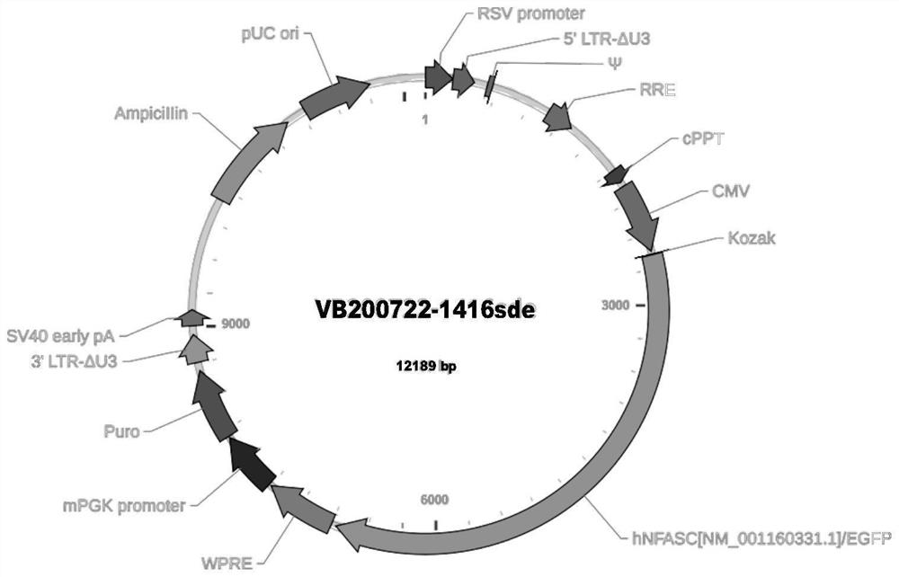
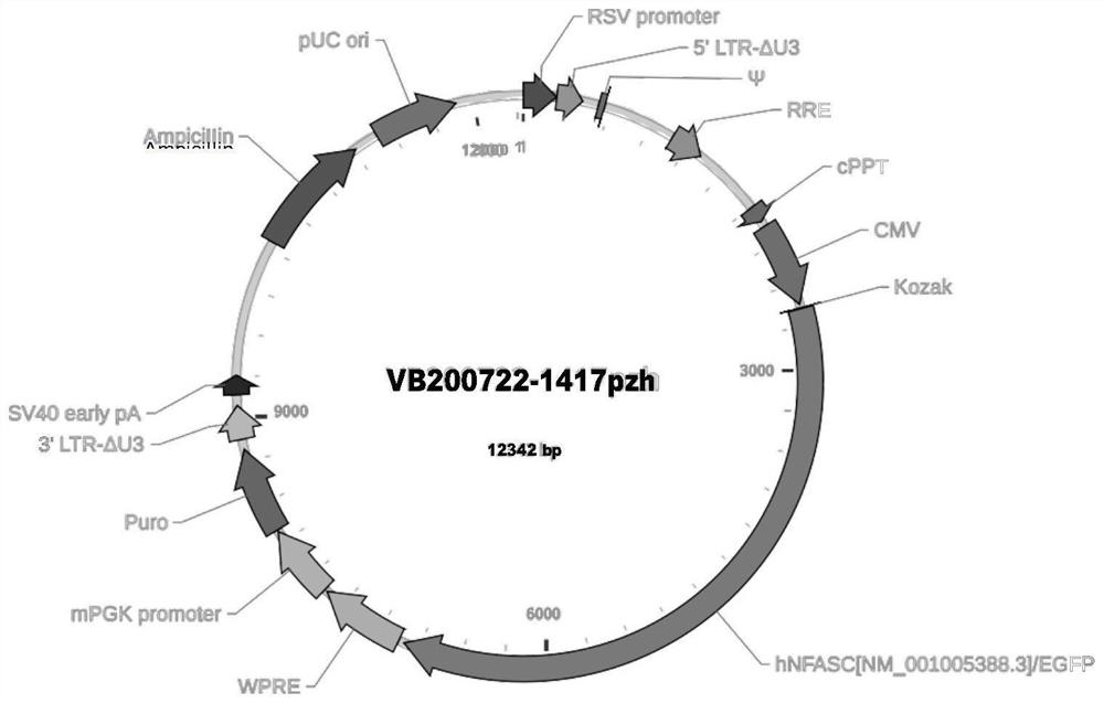
[0024] 1.1 Utilize the homologous recombination method, the target gene NF155 of purification and NF186 are respectively mixed with carrier (such as figure 1 and figure 2 shown) ligation at 50°C for 70 min, and the ligation products were named pLV[Exp]-Puro-CMV>hNFASC[NM_001160331.1] / EGFP and pLV[Exp]-Puro-CMV>hNFASC[NM_001005388.3] / EGFP .

[0025] 1.2 Transform 50 μL supercompetent cells with 10 μL of the ligation product, mix the ligation product with the competent cells, and place on ice for 5 minutes. After heat shock at 42°C for 60 seconds, place it on ice for 1-2 minutes, add 500 μL of LB medium, 100 rpm, 37 Incubate on a constant temperature shaker at ℃ for 30 minutes, centrifuge at 3000 rpm for 2 minutes, discard 400 μL of the culture supernatant, mix the remaining 50-100 μL with a pipette and spread evenly on an LB plate containing 50 μg / mL ampicillin resistance, invert, and keep the temperature at 37 °C Incubate overnight in an incu...

Embodiment 2

[0028] Transfection:

[0029] 2.1 Plate 293T cells: trypsinize the cells and centrifuge and resuspend to plate. The plate requirements are to spread the cells evenly in a six-well plate, and place them in an incubator for overnight culture;

[0030] 2.2 Combine the target plasmid pLV[Exp]-Puro-CMV>hNFASC[NM_001160331.1] / EGFP and pLV[Exp]-Puro-CMV>hNFASC[NM_001005388.3 / EGFP obtained in Example 1 with the packaging plasmid pMDLg / pRRE respectively , PMD2.G, pRSV-Rev, mix according to the ratio of 8:8:2:2; add the plasmid dilution solution to Lipo 2000 dilution solution, mix gently and let stand at room temperature for 10 minutes;

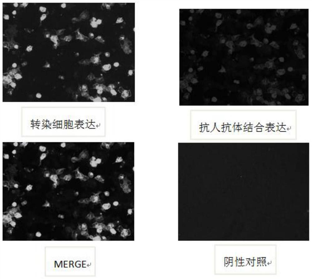
[0031] 2.3 Replace the medium of the six-well plate in 2.1 with 1ml of new complete medium without antibiotics, then add the mixture obtained in 2.2 to each well of the six-well plate and gently shake the cell plate back and forth to make the mixture and Mix the culture solution in the well, culture in a 37°C cell CO2 incubator for 1-3 days, observe t...

Embodiment 3

[0038] divert:

[0039] 3.1 Make the cell density 1-2*10 5 Inoculate the 293T cell suspension per ml into a six-well plate (the confluence of the target cells should be 30%-50% during transduction), and place it in a carbon dioxide incubator at 37°C and 5% CO2 for 18-20 hours;

[0040] 3.2 The initial MOI of the transduced cells is set to 5; thaw the frozen virus solution on ice, mix the thawed virus particles, and use a 6-well plate for transduction; take 10ul>10 8 Add TU / ml virus solution to 1ml culture medium, then mix gently; add 5μg / ml Polybrene at the same time to enhance the binding of pseudovirus capsid and cell membrane; add 0.02g / ml sodium cromoglycate solution at the same time to maintain the cell membrane stability and reduce cell inflammatory response;

[0041]3.3 After 24 hours, suck out the original medium in 3.1, then add the virus-containing medium in 3.2 to the cells, shake the culture plate gently so that the virus liquid can cover every cell, and then pla...


PUM


Abstract

The invention discloses a method for detecting neurotensin NF155 and NF186 antibodies in serum and cerebrospinal fluid, which comprises the following steps: S1, plasmid construction: respectively connecting target genes NF155 and NF186 with a carrier to obtain target plasmids; s2, transfecting 293T cells with the plasmids obtained in the step S1 to obtain viruses; s3, infecting 293T cells with thevirus obtained in the step S2 to obtain transfected cells; and S4, detecting antibodies in serum and cerebrospinal fluid by using the transfected cell expression protein. Proper plasmids are constructed and transfected into cells, a special culture medium is adopted for screening, and after a stable expression effect is generated, whether corresponding antibodies exist or not is accurately detected according to the combination of the corresponding antibodies in serum or cerebrospinal fluid and expression proteins of the antibodies, so that the detection sensitivity and accuracy are improved.

Description

technical field [0001] The invention relates to the technical field of biomedical engineering, in particular to a method for detecting neurofascin NF155 and NF186 antibodies in serum and cerebrospinal fluid. Background technique [0002] Autoantibodies against Ranvier's junction may be an important cause of chronic inflammatory demyelinating polyneuropathy. Possibly a target antigen for autoantibodies. NF belongs to the immunoglobulin superfamily and has two subtypes, NF155 and NF186. NF155 is located in the glial ring in the lateral region of the Ranvier junction, where Caspr and Contactin gather in the lateral region to form septal-like junctions and promote nerve cell adhesion and axon growth. NF186 is located at the junction of Ranvier and axon initiation, where it inhibits cell adhesion and axon outgrowth. Both play an important role in the process of neural development and maintaining the stability of glial cells and axon structures. [0003] At present, the detect...

Claims


Application Information

Patent Timeline
Patent Type & Authority Applications(China)
IPC IPC(8): G01N33/68C07K14/705C12N15/12C12N15/85
CPCC07K14/70503C12N15/85G01N33/68G01N2333/70503
Inventor 周炜滕飞鹏彭确昆
Owner 成都海默云因医学检验实验室有限公司
Why Eureka
Try Eureka