Separating and extracting method for primary liver parenchyma cells and application of primary liver parenchyma cells

A technology of hepatic parenchymal cells and extraction method, which is applied in the field of separation and extraction of primary hepatic parenchymal cells, can solve the problems that liver tissue is difficult to meet the requirements of the two-step perfusion method, reduce the cell yield, etc., and achieve the benefit of adherence and later growth , Good repeatability, and the effect of reducing operation time

Active Publication Date: 2021-01-22
CHINA AGRI UNIV
View PDF5 Cites 4 Cited by
  • Summary
  • Abstract
  • Description
  • Claims
  • Application Information

AI Technical Summary

Problems solved by technology

Liver tissue obtained after feeding experiments in some animals (e.g., sheep) is difficult to meet the requirements of the two-step perfusion method
In addition, the liver tissue of some animals is bulky. If the two-step collagenase perfusion method is completely copied, it...

Method used


Image

  • Separating and extracting method for primary liver parenchyma cells and application of primary liver parenchyma cells
  • Separating and extracting method for primary liver parenchyma cells and application of primary liver parenchyma cells
  • Separating and extracting method for primary liver parenchyma cells and application of primary liver parenchyma cells

Examples


Embodiment 1

[0048] This example provides a method for separating and extracting sheep liver parenchymal cells. details as follows:

[0049] 1) Select a piece of fresh sheep liver tissue with a weight of about 7.5g, and wash the surface of the sheep liver tissue block with 4°C pre-cooled PBS; use a 10mL syringe to puncture the cut surface of the sheep liver tissue with a complete upper surface and a section of blood vessels, The needle was perpendicular to the cut surface, to avoid the needle passing through the tissue block, and the syringe was slowly advanced, and the perfusate Ia (25 mL) preheated at 38°C was injected into the center of the liver tissue, a large amount of blood flowed out, and the liver tissue block in the central area was darkened. The red color gradually turns brownish yellow, and the surrounding tissue blocks are still red in color. Then, at the same angle, inject preheated 38°C perfusate Ib (75mL) into the surrounding tissue area, blood can be seen flowing out, and...

Embodiment 2

[0064] This example provides a method for separating and extracting sheep liver parenchymal cells. The specific method is the same as in Example 1, except that the weight of the liver tissue block is about 15 g, the dosage of perfusate I is 200 mL (50 mL of perfusate Ia, 150 mL of perfusate Ib), and the perfusion time is 15 minutes; the perfusion solution II contains 5.5 mmol / L CaCl 2 and 1.2mg / mL type IV collagenase HBSS solution, the dosage is 300mL, and the perfusion time is 20min.

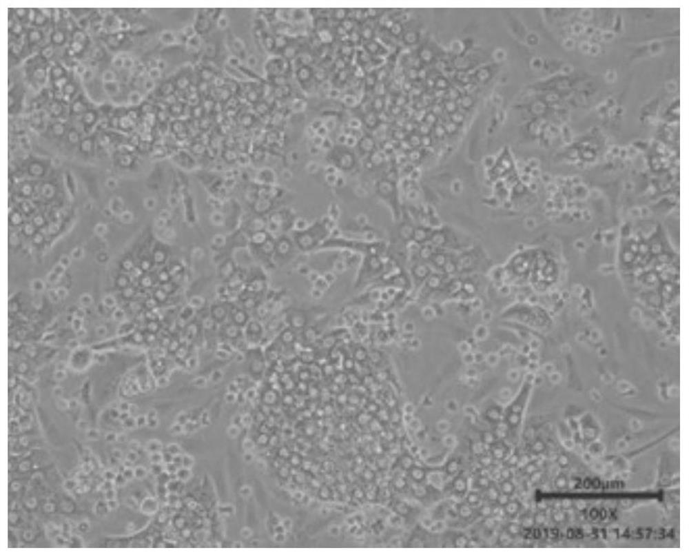
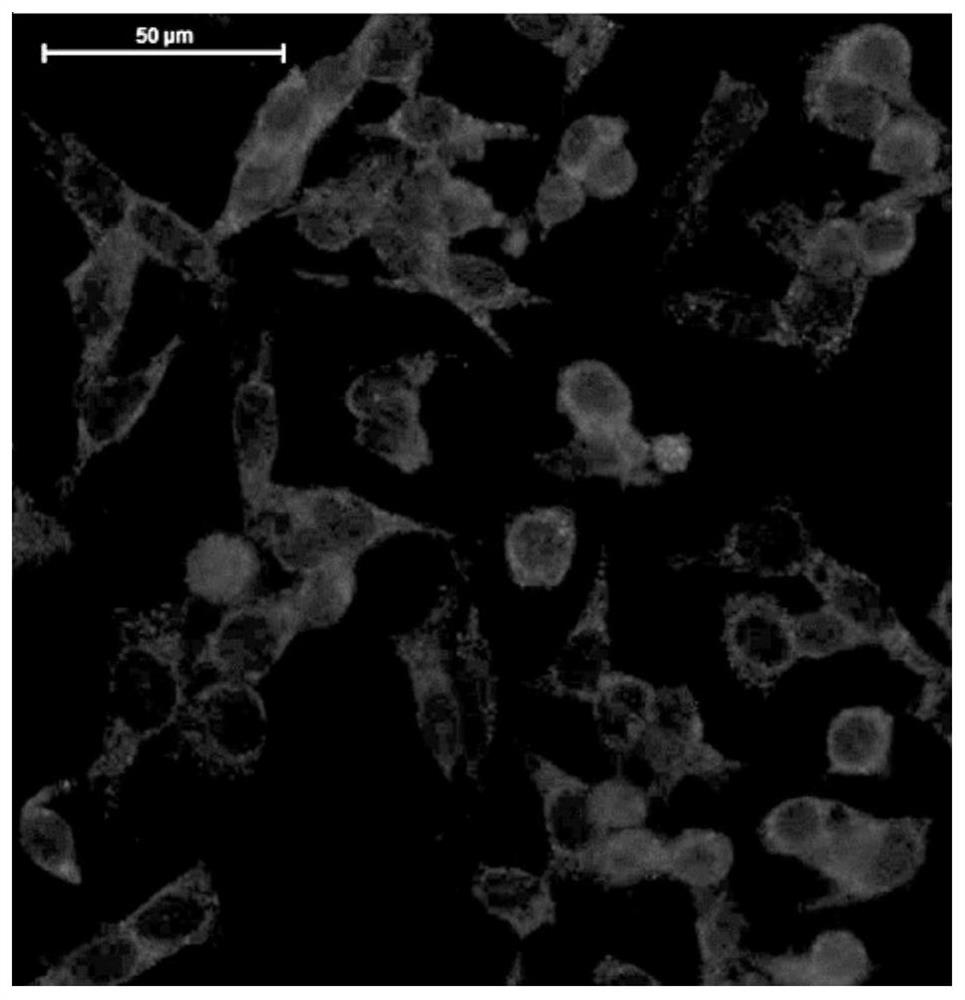
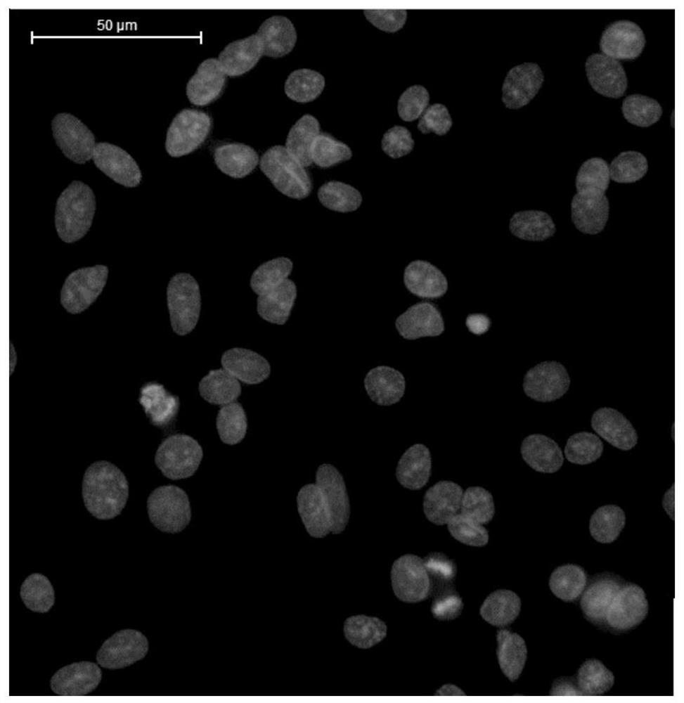
[0065] Experimental results: the cell survival rate reached 95.2%, and the number of living cells was 2.55×10 8 , the cell purity was 96.3%.

Embodiment 3

[0067] This example provides a method for separating and extracting sheep liver parenchymal cells. The specific method is the same as in Example 1, except that the weight of the liver tissue block is about 2.5 g, the amount of perfusate I is 40 mL (7 mL of perfusate Ia, and 33 mL of perfusate Ib), and the perfusion time is 8 minutes; the amount of perfusate II is 50 mL, and the perfusion The time is 6 minutes.

[0068] Experimental results: the cell survival rate reached 94.9%, and the number of living cells was 4.3×10 7 , the cell purity was 95.6%.


PUM


Abstract

The invention relates to the technical field of cell separation and particularly discloses a separating and extracting method for primary liver parenchyma cells and application of the primary liver parenchyma cells. The method disclosed by the invention comprises the steps: injecting perfusate I and perfusate II into a hepatic tissue mass to digest and separate the liver parenchyma cells so as toobtain a cell suspension, then, subjecting filter liquor, obtained after filtering the cell suspension, to normal-temperature centrifugation for 5 times sequentially, wherein centrifugation conditionssequentially are 1000-1580rpm/min*3-5min, 500-650rpm/min*3-5min, 200-300rpm/min*4min, 200-300rpm/min*4min and 200-300rpm/min*4min. According to the method, the operation is simple, consumed time is short, low-temperature centrifugation equipment is not required, red blood cells and cell debris can be efficiently removed, and obtained liver parenchyma cells are relatively high in survival rate andpurity.

Description

technical field [0001] The invention relates to the technical field of cell separation, in particular to a method for separating and extracting primary hepatic parenchymal cells and its application. Background technique [0002] The separation of early hepatic parenchymal cells is generally separated by physical means such as mechanical shearing and strong pipetting, but this method has extremely low cell yield and poor activity. Later, after physically shredding the liver tissue, some researchers added trypsin and collagenase to digest the tissue fragments, which increased the yield of liver parenchymal cells, but the cell survival rate and activity were still not high. Subsequently, the isolated collagenase perfusion method was invented. This method is to take out the animal liver, put a tube in the portal vein or directly put a tube in the blood vessel or incision of the obtained liver tissue block, and use preheated calcium-free and magnesium-free D- Perfuse with Hank's...

Claims


Application Information

Patent Timeline
IPC IPC(8): C12N5/071
CPCC12N5/067C12N2509/00
Inventor 罗海玲简路洋薛瑛
Owner CHINA AGRI UNIV
Why Eureka
Try Eureka