Application of protein SFTPC as lung cancer diagnostic marker and kit

A kit and protein technology, applied in the field of medical diagnosis, can solve the problems of ineffective enrichment of exosomes from tissues or diseases, inability to reflect the specificity of exosomes, complex sources of exosomes, etc., and achieve great theoretical value and clinical application value, good clinical application prospects, and structurally complete effects

Active Publication Date: 2021-03-02
WAYEN BIOTECHNOLGIES SHANGHAI INC
View PDF1 Cites 0 Cited by
  • Summary
  • Abstract
  • Description
  • Claims
  • Application Information

AI Technical Summary

Problems solved by technology

Relatively speaking, the magnetic bead immunocapture method is easy to operate, but currently it is usually based on exosome markers (CD9, CD63) and other exosome membrane proteins, which are generally suitable for the overall capture of exosomes and cannot reflect specific sources. Exosome specificity
Due to the complex sources of exosomes in body fluid samples, the use of body fluid exosomes for disease-related research has the interference of other cell-derived exosomes. For example, in the study of lung cancer exosomes in blood, there will be other tissues, organs, red blood cells Interference with exosomes from other sources, which greatly hinders the research process
Commonly used exosome extraction techniques cannot effectively enrich exosomes from specific tissues or diseases. How to obtain highly specific tissue or disease-derived exosomes is crucial to promote the research and application of exosomes

Method used


Image

  • Application of protein SFTPC as lung cancer diagnostic marker and kit
  • Application of protein SFTPC as lung cancer diagnostic marker and kit
  • Application of protein SFTPC as lung cancer diagnostic marker and kit

Examples


Embodiment 1

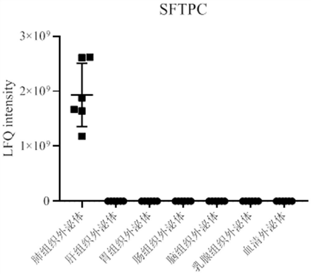
[0066] Example 1: Discovery of the lung tissue exosome surface marker SFTPC

[0067] Methods as below:

[0068] Proteomic analysis of exosomes from different types of tissue sources (lung tissue, liver tissue, gastric tissue, intestinal tissue, brain tissue, breast tissue) and serum exosomes was performed using mass spectrometry. see results figure 1 ,Specific steps are as follows:

[0069] 1. Preparation of tissue exosomes

[0070] (1) Rinse tissue samples with PBS to remove blood stains, remove fat tissue, connective tissue, necrotic tissue and other redundant tissues and the original storage medium;

[0071] (2) Transfer the tissue sample to a petri dish, add an appropriate amount of PBS, and cut the tissue sample with ophthalmic scissors;

[0072] (3) Add collagenase so that the concentration of the working solution is 1 mg / mL, place on a constant temperature shaker at 37°C, 90 rpm, and incubate for about 60 minutes until the tissue block has good light transmission an...

Embodiment 2

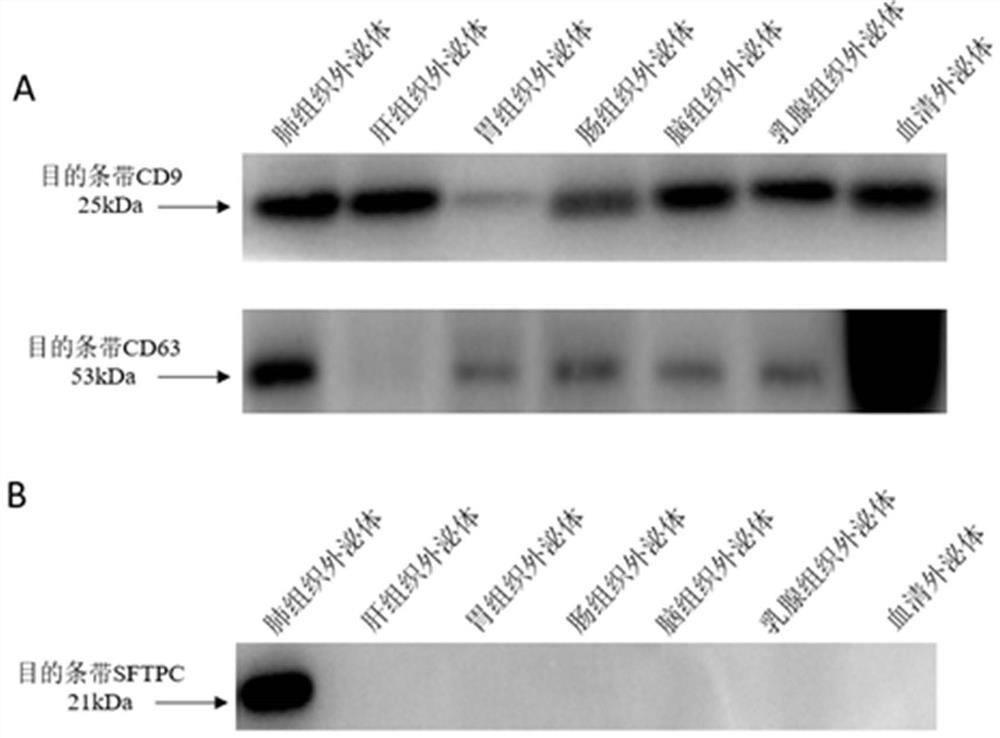
[0120] Example 2: Verification of the lung tissue exosome surface marker SFTPC

[0121] Methods as below:

[0122] Take the PBS resuspension of tissue exosomes and serum exosomes, add an equal volume of RIPA (strong) lysate for lysis and protein extraction, and detect exosome markers and SFTPC by Western blot. see results figure 2 ,Specific steps are as follows:

[0123] (1) Gel preparation: according to the molecular weight of the target protein, use a 1.5 mm glass plate and a 15-hole sample comb to prepare a 10% separating gel and 5% stacking gel;

[0124] (2) Sample loading

[0125] (3) Electrophoresis: conduct electrophoresis at a steady voltage of 80 v until the Loading Buffer (indicator) enters the separation gel, change to a constant voltage of 120 v and continue electrophoresis until the Loading Buffer (indicator) reaches the bottom of the gel, then the electrophoresis can be terminated;

[0126] (4) Membrane transfer: A PVDF membrane with a pore size of 0.22 um w...

Embodiment 3

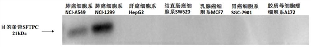
[0134] Example 3: Specific verification of lung tissue exosome surface marker SFTPC

[0135] Methods as below:

[0136] Use the public database Human Protein Atlas (http: / / www.proteinatlas.org / ) and GTEx database (https: / / www.gtexportal.org / home / ) to query the expression of mass spectrometry screening index SFTPC in different tissues, query results It shows that SFTPC is highly specifically expressed only in lung tissue.


PUM


Abstract

The invention provides an application of a quantitative detection agent of protein SFTPC in preparation of a kit for lung cancer diagnosis, auxiliary diagnosis or prognosis analysis, and an application in preparation of a kit for enriching lung-derived exosomes so that the effect of enriching lung tissue specific exosomes can be achieved; the effect of assisting lung cancer diagnosis can also be achieved, and the enriched specific exosome is directly used for detecting lung-related disease markers based on miRNA, protein and the like.

Description

technical field [0001] The invention relates to the field of medical diagnosis, in particular to an application and a kit of protein SFTPC as a diagnostic marker for lung cancer. Background technique [0002] Lung cancer prevention and treatment is a common challenge for China and the world. Screening and early diagnosis and treatment are one of the most effective means to reduce lung cancer deaths. [0003] Extracellular vesicles (EVs) are small membranous vesicles released by different types of cells into the extracellular matrix. Their function is to participate in the transport of molecules between cells, and they exist in almost all major body fluids. EVs contain different types of molecules, including proteins, lipids, DNA, mRNA, miRNA, etc. These content substances will change with the state of parental cells secreting EVs and the changes in their environment, that is, the content substance information carried by EVs Represents the cellular state of the parental cell...

Claims


Application Information

Patent Timeline
Patent Type & Authority Applications(China)
IPC IPC(8): C12N5/071C12Q1/6886G01N33/574
CPCC12N5/0688C12N2509/00C12Q1/6886C12Q2600/158C12Q2600/178G01N33/57423G01N33/57488G01N2800/52
Inventor 张庆华宋凯
Owner WAYEN BIOTECHNOLGIES SHANGHAI INC
Why Eureka
Try Eureka