A kind of self-anastomosis artificial vascular stent and preparation method thereof

A technology of artificial blood vessels and composite stents, applied in the field of biomedical tissue engineering, can solve the problems of long operation time, high quality and technical requirements for surgeons, and high requirements for the operation environment

Active Publication Date: 2022-04-19
SHANGHAI UNIV
View PDF0 Cites 0 Cited by
  • Summary
  • Abstract
  • Description
  • Claims
  • Application Information

AI Technical Summary

Problems solved by technology

[0003] However, in the artificial vascular stent grafting operation, the vascular anastomosis technique of manual suturing is still mainly used at present. This method has a series of unavoidable problems such as long operation time, easy leakage of blood vessels, and high requirements for the operation environment. High self-quality and technical requirements
In the prior art, in order to avoid the above problems, although many non-suture techniques have appeared, such as magnetic tube method, vascular anastomosis clip, needle loop method, vascular adhesive, etc., these non-suture techniques usually require a large number of auxiliary tools Vascular anastomosis can only be completed, and the anastomotic effect is not good

Method used


Image

  • A kind of self-anastomosis artificial vascular stent and preparation method thereof
  • A kind of self-anastomosis artificial vascular stent and preparation method thereof
  • A kind of self-anastomosis artificial vascular stent and preparation method thereof

Examples


preparation example Construction

[0028] The invention provides a method for preparing a self-anastomotic artificial vascular stent, comprising the following steps:

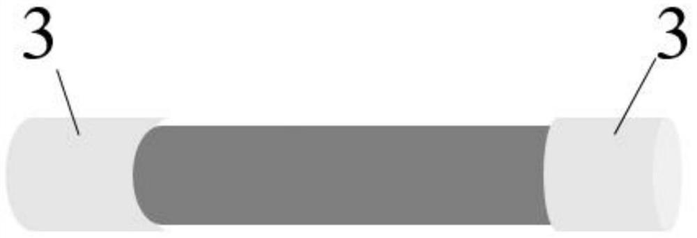
[0029] (1) Using the polymer solution as the spinning solution, the electrospun membrane is prepared by electrospinning, and then the electrospun membrane is rolled to obtain the vascular inner layer stent;

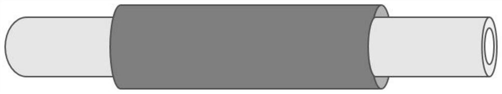
[0030] (2) Using a mixed solution of sodium alginate, gelatin and carbon nanotubes as the printing material, extruding and printing the middle layer of the blood vessel bracket on the outer surface of the vascular inner layer stent obtained in the step (1), to obtain a double-layer stent;

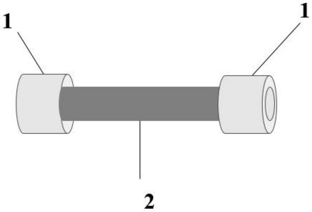
[0031] (3) Using shape memory material as the printing material, extrude and print an anastomotic sleeve at both ends of the double-layer stent in the step (2) to form a composite stent; the inner diameter of the anastomotic sleeve and the inner layer of the blood vessel The inner diameter of the bracket is the same;

[0032] (4) Using polycaprolactone s...

Embodiment 1

[0083] a. Preparation of endovascular stent

[0084] Configuration of polymer solution:

[0085] (1) Prepare a 5% heparin solution with 0.05mol / L, pH=6.0 2-morpholineethanesulfonic acid buffer solution;

[0086] (2) Mix 1-(3-dimethylaminopropyl)-3-ethylcarbodiimide hydrochloride, N-hydroxysuccinimide and the above-mentioned heparin solution according to the ratio of 2:1.3:1 Mix and react for 30 minutes to obtain a mixed solution; then add 1 g of silk fibroin to 50 mL of the above mixed solution, and obtain a heparinized silk fibroin solution after 5 hours;

[0087] (3) Take 2g of polycaprolactone and 10mL of heparinized silk fibroin solution and dissolve them in 10mL of hexafluoroisopropanol to obtain a polymer solution;

[0088] Preparation of vascular inner layer scaffold by electrospinning:

[0089] Put the above polymer solution in a water tank at 37°C, stir at a rate of 400r / min for 0.5h, let it stand for 1h, then put it into a syringe, select a 23G nozzle, set the hig...

Embodiment 2

[0109] a. Preparation of endovascular stent

[0110] Configuration of polymer solution:

[0111]Take 2 g of polycaprolactone, dissolve it in a mixed solvent made of 7 mL of dichloromethane and 3 mL of dimethylformamide, and let it stand for 6 hours to fully dissolve the polycaprolactone to obtain a polycaprolactone solution;

[0112] Preparation of vascular inner layer stent:

[0113] Stir the obtained polycaprolactone solution for 0.5h, let it stand for 1h, defoam, put it into a syringe, select a 23G nozzle, set the high-voltage voltage parameter to 10kV, and set the distance from the nozzle to the collector to 10cm, and then use 16.6μL Electrospun at a flow rate of / min to obtain an electrospun membrane; the obtained electrospun membrane was rolled into a tubular shape with a stainless steel rod with a diameter of 4 mm, and 8 cm was selected as the inner layer of the blood vessel to obtain an inner diameter of 4 mm, a length of 8 cm, and a thickness of 4 mm. 0.2mm vascular...


PUM

PropertyMeasurementUnit
thicknessaaaaaaaaaa
lengthaaaaaaaaaa
thicknessaaaaaaaaaa

Abstract

The invention provides a method for preparing a self-anastomating artificial vascular stent. A three-layer artificial vascular stent similar to the structure of a natural blood vessel is prepared by electrospinning and extrusion printing, and a shape memory material is used on the inner layer of the vascular stent and the blood vessel. The two ends of the double-layer stent composed of the middle layer stent are extruded and printed with an anastomotic sleeve with the same inner diameter as the vascular inner layer stent, and then the deformation of the anastomotic sleeve can be controlled by changing the external temperature, realizing the artificial blood vessel without auxiliary tools and The purpose of self-anastomosis between sutures and autologous blood vessels. The results of the examples show that after inserting the anastomotic sleeve of the self-anastomating artificial vascular stent prepared by the present invention into the broken end of the autologous blood vessel, and then heating it, the diameter of the broken end of the autologous blood vessel will become larger under the support of the anastomotic sleeve, thereby achieving autologous The purpose of anastomosis of blood vessels and self-anastomotic artificial vascular stents without auxiliary tools.

Description

technical field [0001] The invention relates to the technical field of biomedical tissue engineering, in particular to a self-anastomizing artificial blood vessel stent and a preparation method thereof. Background technique [0002] Cardiovascular disease is the cause of the highest mortality rate in the world, and vascular bypass grafting is considered to be the best choice for patients who need long-term maintenance of revascularization, among which autologous blood vessels (such as saphenous vein, radial artery and internal thoracic artery) However, due to the lack of sources of autologous blood vessels and the trauma damage to the cut site, the wide application of autologous blood vessel transplantation is greatly limited. Based on this, people have researched and developed artificial vascular stents as an ideal substitute for autologous blood vessels. [0003] However, in the artificial vascular stent grafting operation, the vascular anastomosis technique of manual sut...

Claims


Application Information

Patent Timeline
Patent Type & Authority Patents(China)
IPC IPC(8): G06F30/00A61F2/07A61L27/08A61L27/18A61L27/20A61L27/22A61L27/50A61L27/40
CPCA61F2/07A61L27/20A61L27/222A61L27/08A61L27/18A61L27/50A61L27/58A61L2400/16A61L2400/12C08L5/04C08L67/04
Inventor 胡庆夕王琪张海光
Owner SHANGHAI UNIV
Why Eureka
Try Eureka