Application of MYOG gene as target point in preparation of drugs for treating cardiovascular diseases related to myocardial cell apoptosis

A technique for cardiomyocyte apoptosis and cardiovascular application in the field of biomedicine

Active Publication Date: 2021-04-06
GUANGDONG BEATING ORIGIN REGENERATIVE MEDICINE CO LTD
View PDF2 Cites 3 Cited by
  • Summary
  • Abstract
  • Description
  • Claims
  • Application Information

AI Technical Summary

Problems solved by technology

However, the expression of MYOG gene in human heart tissue a

Method used


Image

  • Application of MYOG gene as target point in preparation of drugs for treating cardiovascular diseases related to myocardial cell apoptosis
  • Application of MYOG gene as target point in preparation of drugs for treating cardiovascular diseases related to myocardial cell apoptosis
  • Application of MYOG gene as target point in preparation of drugs for treating cardiovascular diseases related to myocardial cell apoptosis

Examples


Embodiment 1

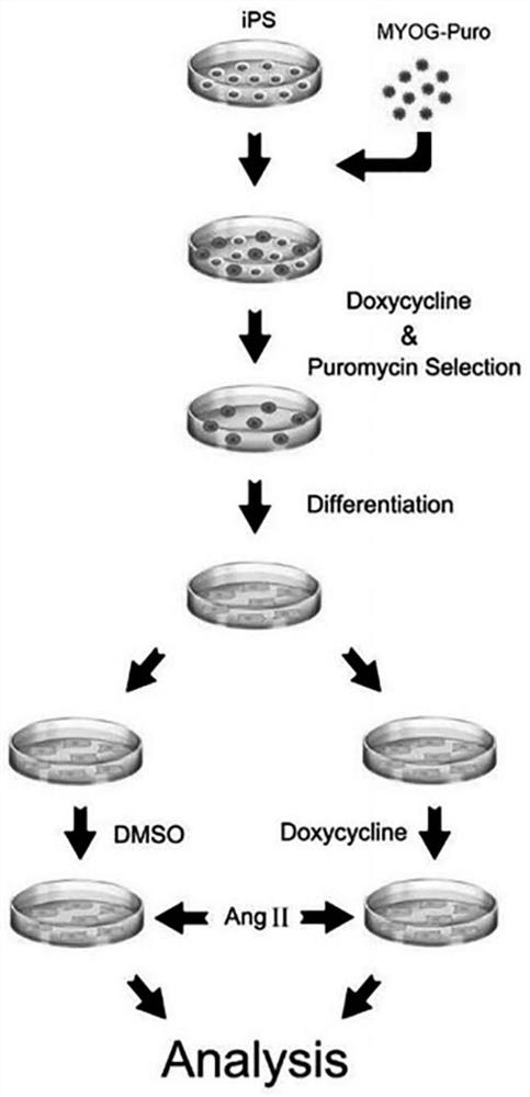
[0029] Example 1: Obtaining hIPSC-MYOG cell lines

[0030] 1.1 Construction of lentiviral expression vector: MYOG cDNA and a puromycin resistance gene were subcloned into pCW-Cas9-Blast vector (Addgene, 83481) by conventional molecular cloning methods, and Cas9 and Blast genes in the original vector were replaced to obtain pCW-MYOG.

[0031] 1.2 Lentiviral packaging

[0032] 1.2.1 Inoculate HEK293T cells into a 6-well plate, culture with D10 medium (DMEM medium + 10% fetal calf serum), and prepare for transfection when the cell confluence reaches 70%-80%.

[0033] 1.2.2 One hour before transfection, the original culture medium was discarded, and 2 mL / well of preheated serum-free OptiMEM culture medium was added.

[0034] 1.2.3 Perform transfection with Lipofectamine 2000 reagent according to the instructions. HEK293T cells were co-transfected with pCW-MYOG (20 μg), pVSVg (10 μg) (Addgene), and psPAX2 (15 μg) (Addgene).

[0035] 1.2.4 After 6 hours, replace the culture medi...

Embodiment 2

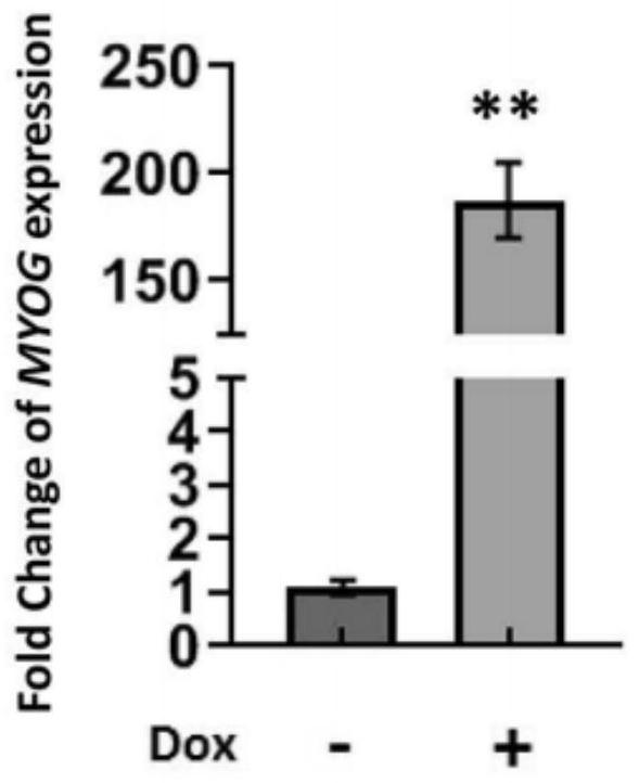
[0042] Example 2: Dox induces MYOG expression

[0043]2.1 Induction: Add doxycycline hydrochloride (Dox) (Sigma, D9891) at a final concentration of 2 μg / mL to STEMUP to induce the expression of MYOG, and use DMSO as a control. The control group has no DOX but only DMSO, marked as C1, and added to STEMUP; the test group has both DOX and DMSO, and DOX is first added to DMSO, marked as C2, and then added to STEMUP; the final concentration of DOX in STEMUP is 2 μg / mL; C1 The same as the addition of C2;

[0044] 2.2 Total RNA extraction: Total cellular RNA was extracted with UNlQ-10 Column Trizol Total RNA Extraction Kit (Sangon Biotech, B511321-0100). (The sample was treated with DNase I (deoxyribonuclease I, Sangon Biotech, B618252) for 30 minutes in advance);

[0045] 2.3 Reverse transcription: RNA was reverse-transcribed using the reverse transcription kit iScript Reverse Transcription Supermix (Bio-Rad, 1708841).

[0046] 2.4 qPCR detection of MYOG mRNA expression level: ac...

Embodiment 3

[0052] Example 3: Obtaining hiPSC-CM / hiPSC-MYOG-CM

[0053] 3.1 The differentiation of hiPSC: In RPMI-BSA medium [RPMI1640 medium (HyClone, SH30027.01) + 213 μg / mL AA2P (l-ascorbic acid 2-magnesium phosphate) (Sigma, A8960) and 0.1% bovine serum albumin (Sigma , A1470)], add the small molecule CHIR99021 (Tocris, 4423, the final concentration is 10mM)] to treat hiPSCs for 24 hours, and then change the RPMI-BSA medium and incubate for 48 hours. On the 4th day of differentiation, the small molecule IWP2 (Tocris, 3533, final concentration 5 μM) was added to the RPMI-BSA medium to treat the cells. After 48h, the medium was replaced with RPMI-BSA medium. At this stage, it differentiates from hipsc to hipsc-cm. In subsequent experiments, cardiomyocytes were cultured with RPMI1640 medium plus 3% serum replacement (Gibco, 10828-028).

[0054] 3.2 Purification of hiPSC-MYOG-CM: hiPSC-CM was purified using a metabolic selection method. The metabolic selection medium was DMEM medium (...


PUM


Abstract

The invention belongs to the technical field of biological medicines, and particularly relates to application of an MYOG gene as a target point in preparation of drugs for treating cardiovascular diseases related to myocardial cell apoptosis. According to the invention, an angiotensin II induced human induced pluripotent stem cell differentiation myocardial cell apoptosis model is constructed in vitro, the effect of a transcription factor MYOG in inhibiting myocardial cell apoptosis is disclosed for the first time, and a theoretical basis and a scientific basis are provided for research and development of drugs for cardiovascular diseases related to myocardial cell apoptosis, thereby being a new target for research and development of cardiovascular disease drugs.

Description

technical field [0001] The invention belongs to the technical field of biomedicine, and specifically relates to the application of the MYOG gene as a target in the preparation of drugs for treating cardiomyocyte apoptosis-related cardiovascular diseases. Background technique [0002] Cardiovascular disease (CVD) is currently the number one killer of human health, and the number of people dying from cardiovascular disease is more than any other reason in the world every year. According to the statistics of the World Health Organization (WHO), more than 17 million people worldwide die from CVD each year, accounting for 31% of the total global deaths. With the improvement of people's living standards and changes in dietary structure, the prevalence and mortality of cardiovascular diseases have shown a clear upward trend. [0003] Cardiomyocyte apoptosis is involved in the pathophysiological process of various cardiovascular diseases, including essential hypertension, ischemic ...

Claims


Application Information

Patent Timeline
IPC IPC(8): C12Q1/02C12N15/867C12N5/10
CPCG01N33/5061C12N15/86C12N5/0657C07K14/4705C12N2800/107C12N2740/15043C12N2503/02C12N2506/45C12N2510/00G01N2500/10G01N2500/04C12N2740/16043A61P21/00G01N2333/4706
Inventor 曾嵘林彬
Owner GUANGDONG BEATING ORIGIN REGENERATIVE MEDICINE CO LTD
Why Eureka
Try Eureka