Fusion protein and application thereof in preparation of novel coronavirus subunit vaccine

A fusion protein and virus technology, applied in the field of vaccines, can solve the problems of weak immunogenicity and enhanced antibody-mediated diseases, and achieve the effect of prolonging the half-life

Pending Publication Date: 2021-05-28
ZHEJIANG UNIV +1
View PDF0 Cites 4 Cited by
  • Summary
  • Abstract
  • Description
  • Claims
  • Application Information

AI Technical Summary

Problems solved by technology

However, full-length S protein vaccines may be able to induce unwanted immune responses leading to enhanced a

Method used


Image

  • Fusion protein and application thereof in preparation of novel coronavirus subunit vaccine
  • Fusion protein and application thereof in preparation of novel coronavirus subunit vaccine
  • Fusion protein and application thereof in preparation of novel coronavirus subunit vaccine

Examples


Embodiment 1

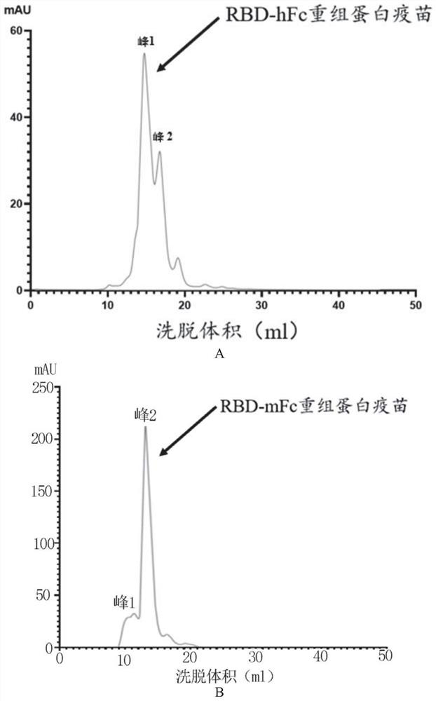
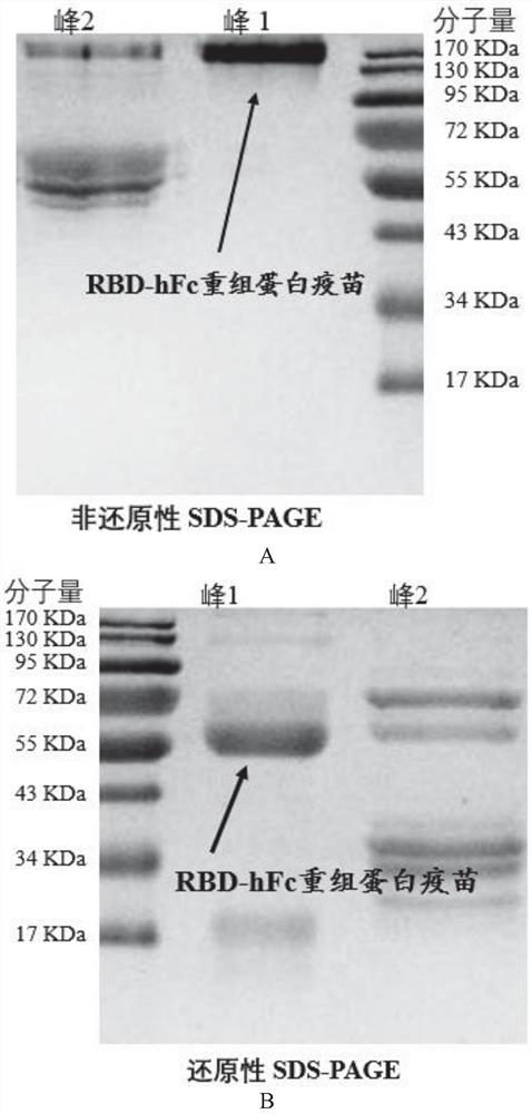
[0026]Example 1: Preparation of RBD recombinant protein

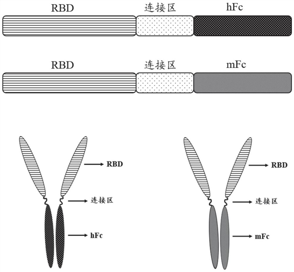
[0027](1) The RBD sequence is derived from the SARS-COV-2 penetration protein 330-583 amino acid range. In the present invention, the source Fc from the mouse source IgG1, the human source Fc is the transformation on the basis of human IgG1. The specific transformation site of human source FC is in ASN297ala and Lys322ala, and the ADCC and CDC effects mediated by FC are redundant. In order to increase the safety of the vaccine, the two sites are mutated.

[0028]Experimental Design Route: First PCD (Gene Sequence SEQ ID No.4, Amino Acid Sequence SEQ ID NOT), and Human IgG1 modified Fc fragment (hereinafter simply referred to as HFC, gene sequence SEQ ID NO) 5, amino acid sequence SEQ ID No. 2), the Fc fragment (hereinafter simply referred to as MFC, gene sequence SEQ ID NO. 6, amino acid sequence SEQ ID NO.). By overlapping the PCR, RBD and HFC and MFC are spliced ​​through a flexible connection zone (Gly-Gly-Gly-ser, gene sequence...

Embodiment 2

[0031]Example 2: Flow affinity determination

[0032](1) Collection 1 × 10⁶VERO E6 cells were respectively composed of RBD-HFC recombinant protein vaccine (10 μg / ml), RBD-MFC recombinant protein vaccine (10 μg / ml), and two groups of negative controls (only PBS only). It was incubated at 4 ° C for 30 min.

[0033](2) After washing of PBS was 3 times, the second anti-rats of Fitc labeled goats were added to the Test tube of RBD-MFC; and the addition of FITC labeled goats were added to the test tube of RBD-MFC. Second Anti-(1: 1000) 200 μL; the negative control group, added to the Fitc labeled goat anti-human secondary antibody (1: 1000) 200 μL, FITC-labeled goat anti-mouse two-resistance (1: 1000) 200 μL. After incubation of 4 ° C for 30 min, PBS was washed 3 times, and the flow cytometer was detected.

[0034](3) Flow cytometry detection of affinity of RBD recombinant protein and Vero E6 cells (ACE2 positive) is mainly expressed by the average fluorescence intensity of FITC after binding....

Embodiment 3

[0036]Example 3: Recombinant protein immunization mice

[0037]Choosing healthy BALB / C female mice, 8 weeks (19-21g), mice were randomly divided into 7 groups, and 4 of each group, F, G, H, I, J, K, L total 7 groups, Specifically, the F group is inoculated by Al (OH)3Adjurant group; Group g is a high dose (8 μg) RBD-HFC (RBD-HFC)HIGH ), H group is a low dose (2 μg) RBD-HFC (RBD-HFC)Low); Group K is inoculated with high dose (8 μg) RBD-MFC (RBD-MFC)HIGH ), Group L is a low dose (2 μg) RBD-MFC (RBD-MFC)Low). Further, in order to compare the immunogenic differences in the recombinant protein vaccine in the two Fc fusion forms of RBD and S1, S1 recombinant protein vaccine fusion human source Fc is provided as a control, S1-HFC (purchased from Beijing Yiqiao Shenzhou Company, Item No. 40591-V02H) In the S1 domain selection S protein 16-685AA, where the Fc is a human source IgG1 (ASN297, and the Lys322 are not mutated).

[0038]Specific as follows: I group is inoculated with high dose (8 μg) ...


PUM


Abstract

The invention discloses a fusion protein and application thereof in preparation of a novel coronavirus subunit vaccine. The fusion protein comprises an RBD region of SARS-CoV-2 virus S protein and an Fc region of an IgG antibody, wherein the RBD region and the Fc region are subjected to fusion expression. A recombinant fusion protein is obtained by performing fusion expression on an RBD structural domain of a spinous process protein of SARS-CoV-2 and an immunoglobulin Fc, and a hinge region and a constant region of the Fc can combine two heavy chains of an IgG antibody through a disulfide bond, so that an RBD dimer is formed; meanwhile, the prepared recombinant fusion protein can be used as a recombinant protein vaccine aiming at novel coronavirus pneumonia. The recombinant protein vaccine can induce generation of a high-titer specific IgG antibody and a neutralizing antibody in a mouse body, the antibody level can be maintained for more than 3 months, and the vaccine can induce the mouse to generate cellular immunity at the same time.

Description

Technical field[0001]The present invention relates to the field of vaccine, and more particularly to a fusion protein and its application in the preparation of new coronavirus subunit vaccines.Background technique[0002]The coronavirus belongs to the Nitro virus, a coronary virus, a single stranded RNA virus (+ SS RNA) having an envelope of the film, and can be divided into α, β, γ, δ genus. Among them, α and β are mainly infected with mammals, while γ, δ is mainly infected with poultry, and in a small number of mammals. The new coronavirus pneumonia (COVID-19) is caused by the SARS-COV-2 virus of the beta.[0003]SARS-COV-2 virus whose genome encodes four structural proteins, respectively, of spine protein, envelope protein, membrane protein, and nuclei protein. Among them, the receptor binding domain of the s protein is infected with the human body by binding to the blood vessel transformase 2 (ACE2) on the host cell membrane. In the absence of a special medicine, the vaccine is stil...

Claims


Application Information

Patent Timeline
IPC IPC(8): C07K19/00C12N15/62A61K39/215A61P31/14
CPCA61K39/12A61P31/14C07K14/005C07K2319/30C12N2770/20022C12N2770/20034
Inventor 周展周晶晶蒋健敏朱函坪孙一晟姚萍萍陈晨赵文彬陈枢青
Owner ZHEJIANG UNIV
Why Eureka
Try Eureka