Construction method and application of retinal vasculopathy model

A technology of retinal blood vessels and construction methods, applied in the field of eye disease research, can solve problems such as lack of animal models, and achieve the effect of assisting research and development and enriching theoretical foundations

Active Publication Date: 2021-06-18
SICHUAN PROVINCIAL PEOPLES HOSPITAL
View PDF8 Cites 3 Cited by
  • Summary
  • Abstract
  • Description
  • Claims
  • Application Information

AI Technical Summary

Problems solved by technology

Currently, there is a lack of animal models available for RNV therapeutic research

Method used


Image

  • Construction method and application of retinal vasculopathy model
  • Construction method and application of retinal vasculopathy model
  • Construction method and application of retinal vasculopathy model

Examples


Embodiment 1

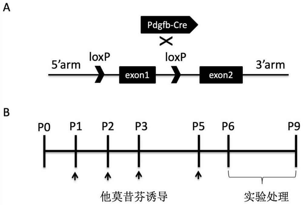
[0054] In this example, mice were used as the target animals, and the Cre-loxp conditional knockout system was used to construct a mouse model of retinal neovascular disease. Tool mice were purchased from University College London, London, UK. Jup carrying the loxp sequence loxp / loxp Mice were purchased from Jackson Laboratory, USA (https: / / www.jax.org / strain / 017575).

[0055] The methods used to build the model include:

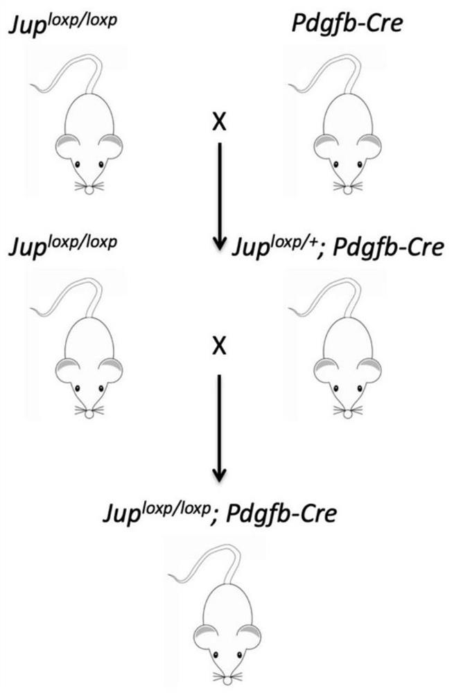
[0056] (1) mice carrying the loxp sequence (Jup loxp / loxp ) was mated with a tool mouse carrying Pdgfb-Cre to obtain a mouse carrying both the loxp sequence and Cre, expressed as Jup loxp / + -Pdgfb-Cre, followed by Jup loxp / loxp with Jup loxp / + -Pdgfb-Cre mating to obtain Jup loxp / loxp -Pdgfb-Cre mice (refer to figure 1 As shown in A, the Cre-loxp system was used to knock out the second exon of Jup in mouse vascular endothelial cells, figure 2 The breeding roadmap of Jup vascular endothelial cell conditional knockout mice is shown in ). ...

Embodiment 2

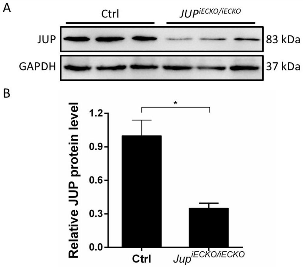
[0060] In this example, western blotting was used to detect the expression of JUP protein in the lung tissue of the target animal on day 25 after birth.

[0061] (1) Harvest the lung tissues of littermate wild-type and knockout-type target mice, put them into 1×PBS containing protease inhibitors, and ultrasonically lyse them on ice for 10 minutes; centrifuge at 16000g for 10 minutes at 4°C, and take Transfer the supernatant to another clean centrifuge tube, add protein loading buffer, mix well and heat at 95°C for 5min.

[0062] (2) After the samples were cooled, 20 μl of protein samples were taken respectively, and electrophoresis was performed at a constant voltage of 70 v for 25 minutes and at 160 v for 40 minutes with 10% separating gel.

[0063] (3) The membrane transfer conditions were as follows: transfer at a constant flow of 0.3A for 1 hour and 30 minutes, then wash the membrane once with deionized water, and block with 5% skimmed milk blocking solution at roo...

Embodiment 3

[0068] In this embodiment, the development of retinal blood vessels is detected by retinal wholemount technology.

[0069] After the mice were killed, the eyeballs were fixed in 4% PFA solution at room temperature for 20 minutes (a 24-well plate can be used as a container), and then soaked in 1×PBS for 15 minutes. Then use a 30G needle to make a hole in the center of the cornea, cut off the entire cornea with ophthalmic scissors, and take out the crystal. Then the whole sclera was torn apart with tweezers, leaving the inner retina, and cut into a four-leaf clover shape, paved to remove excess tissue, inhaled 4% PFA, and continued to fix at 4°C for 24 hours. The wholemount was transferred to 0.4% PFA the next day and stored at 4°C.

[0070] Wholemount staining method: take out the wholemount, wash with 1×PBS 3 times, 5 minutes each time. Subsequently, 100 μL of blocking solution (5% fetal bovine serum, 0.3% TritonX-100, 0.03% sodium azide in PBS) was used to block for 1...


PUM

PropertyMeasurementUnit
Thicknessaaaaaaaaaa

Abstract

The invention discloses a construction method and application of a retinal vasculopathy model, and relates to the field of eye disease research. The Jup gene in vascular endothelial cells of a target animal is not expressed or is inhibited in expression through a genetic engineering technique, so that the target animal shows typical retinal neovascularization disease characteristics. The target animal can be used as a retinal neovascularization disease model for research of the retinal neovascularization disease, can also help to clarify the pathogenesis process and mechanism of the retinal neovascularization disease and enrich the theoretical basis of retinal neovascularization, and provides a new target for treatment or prevention of the retinal neovascularization disease to assist research and development of retina neovascularization treatment means.

Description

technical field [0001] The invention relates to the field of eye disease research, in particular to a method for constructing a retinal vascular disease model and its application. Background technique [0002] At present, apart from laser photocoagulation and intraocular injection of anti-vascular endothelial growth factor (VEGF) drugs in the early stage of the disease, there is no more effective clinical treatment for retinal neovascularization (RNV). treatment method. Existing studies have shown that anti-VEGF therapy is not effective for all patients. At the same time, anti-VEGF therapy has many disadvantages such as high price, repeated injections, and increased risk of infection in patients, which limits its clinical application. Therefore, while conducting research on the pathogenesis of RNV, it is an urgent medical problem to construct effective animal models to seek new effective treatments or therapeutic targets. Currently, there is a lack of animal models availab...

Claims


Application Information

Patent Timeline
IPC IPC(8): C12N15/85C12N15/12A01K67/027
CPCC12N15/8509C07K14/47A01K67/0276A01K2217/075A01K2267/03
Inventor 杨正林朱献军李姝锦杨牧何韵琪
Owner SICHUAN PROVINCIAL PEOPLES HOSPITAL
Why Eureka
Try Eureka