Bone segmentation method in hip joint image, electronic equipment and storage medium

A technology in hip joints and images, applied in the field of image processing, can solve problems such as easy overfitting, unrobust training process, and unfavorable for dense prediction tasks, etc., and achieve the effect of robust results and fast calculation speed

Active Publication Date: 2021-06-22
刘慧烨 +1
View PDF7 Cites 3 Cited by
  • Summary
  • Abstract
  • Description
  • Claims
  • Application Information

AI Technical Summary

Problems solved by technology

[0003] (1) A large number of convolutional layers and downsampling layers reduce the resolution of feature maps, which is not conducive to dense prediction tasks like image segmentation;
[0004] (2) The very deep network structure makes the training process unrobust, easy to overfit, and requires a large amount of data for learning;
[0005] (3) The computational complexity is high, and the prediction speed of the network is limited

Method used


Image

  • Bone segmentation method in hip joint image, electronic equipment and storage medium
  • Bone segmentation method in hip joint image, electronic equipment and storage medium
  • Bone segmentation method in hip joint image, electronic equipment and storage medium

Examples


Embodiment 1

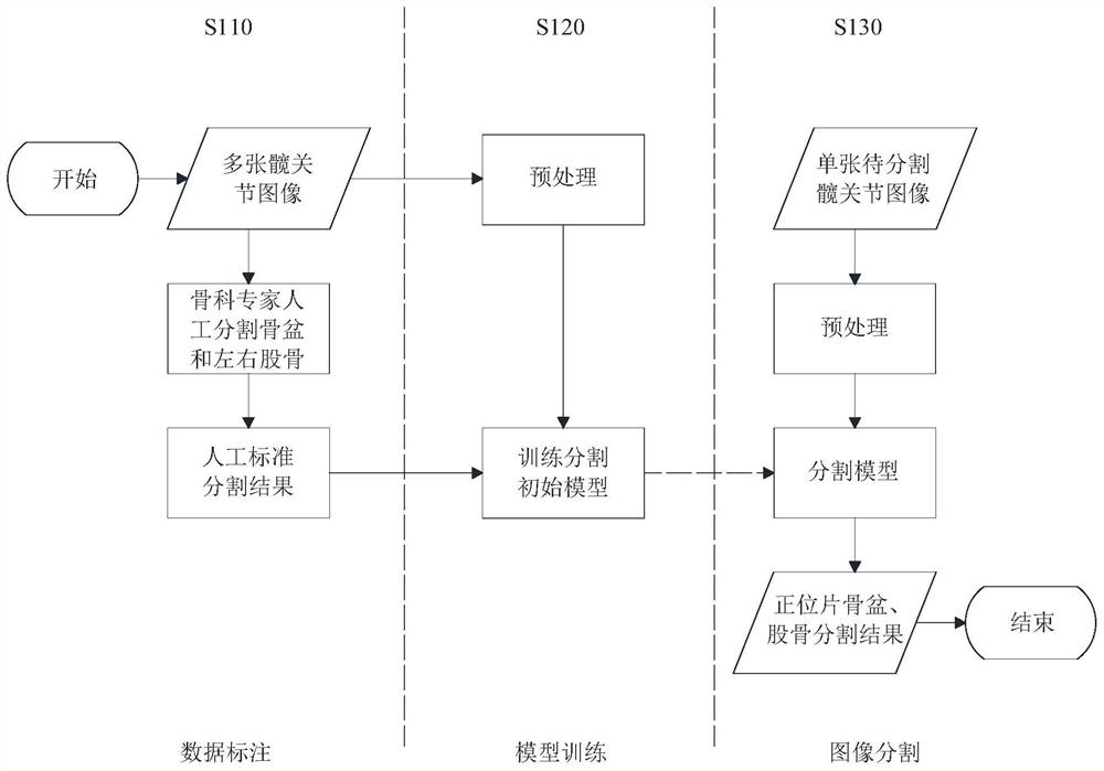
[0076] see figure 1 , figure 1 This is a schematic flowchart of a method for segmenting bones in a hip joint image disclosed in an embodiment of the present invention. Wherein, the execution body of the method described in the embodiment of the present invention is an electronic device composed of software or / and hardware, and the execution body can receive relevant information in a wired or / and wireless manner (mainly receiving hip joint sample images during training). , during the segmentation, the image of the hip joint to be segmented is mainly received), in some embodiments, it may also send certain instructions, and may also have certain storage functions. The execution body may be a computer or server with certain processing functions, and the server may be a physical server or a cloud server. Of course, if the processing capability is sufficient, the execution body may also be a mobile phone or a tablet computer. like figure 1 As shown, the bone segmentation method ...

Embodiment 2

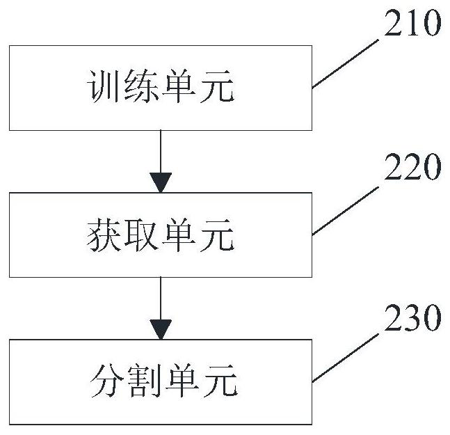
[0102] see image 3 , image 3 It is a schematic structural diagram of a bone segmentation device in a hip joint image disclosed in an embodiment of the present invention. like image 3 As shown, the bone segmentation device in the hip joint image may include:

[0103] The training unit 210 is used for training to obtain a segmentation model.

[0104] an acquisition unit 220, configured to acquire an image of the hip joint to be segmented;

[0105] A segmentation unit 230, configured to input the to-be-segmented hip joint image into a pre-trained segmentation model, to output the segmentation result of the to-be-segmented hip joint image;

[0106] Among them, please refer to Figure 4 As shown, the training unit 210 includes:

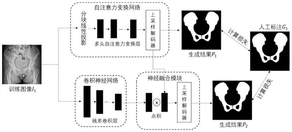
[0107] The creation subunit 211 is used to create a segmentation initial model, and the segmentation initial model includes a self-attention transformation initial model and a convolutional neural network initial model;

[0108] The labeling subu...

Embodiment 3

[0137] see Figure 5 , Figure 5 It is a schematic structural diagram of an electronic device disclosed in an embodiment of the present invention. The electronic device may be a computer, a server, etc. Of course, under certain circumstances, it may also be a smart device such as a mobile phone, a tablet computer, and a monitoring terminal. like Figure 5 As shown, the electronic device may include:

[0138] a memory 310 storing executable program code;

[0139] a processor 320 coupled to the memory 310;

[0140] The processor 320 invokes the executable program code stored in the memory 310 to execute some or all of the steps in the method for segmenting bones in a hip joint image in the first embodiment.

[0141] An embodiment of the present invention discloses a computer-readable storage medium storing a computer program, wherein the computer program causes a computer to execute some or all of the steps in the method for segmenting bones in a hip joint image in the firs...


PUM


Abstract

The embodiment of the invention relates to the field of image processing, and discloses a bone segmentation method in a hip joint image, electronic equipment and a storage medium. The method comprises the steps: obtaining a to-be-segmented hip joint image; inputting the to-be-segmented hip joint image into a pre-trained segmentation model, and outputting a segmentation result, wherein a method for obtaining the segmentation model through pre-training comprises the following steps: creating an initial segmentation model, and obtaining a plurality of artificially labeled hip joint sample images to obtain a mask image; inputting the hip joint sample images into a self-attention transformation initial model and a convolutional neural network initial model to respectively obtain a first segmentation result and a second segmentation result; and calculating training loss, and returning the training loss to the initial segmentation model to obtain a final segmentation model. According to the embodiment of the invention, the segmentation result is accurate, the robustness is realized, and bone structures in the hip joint images can be efficiently and automatically segmented, so that clinical doctors are assisted in surgical planning, intraoperative navigation and postoperative evaluation.

Description

technical field [0001] The invention relates to the technical field of image processing, in particular to a bone segmentation method in a hip joint image, an electronic device and a storage medium. Background technique [0002] The segmentation of hip joint images has always been one of the hotspots in medical imaging research. Accurate and stable segmentation results can well assist orthopaedic surgeons in diagnosing hip joint diseases, quickly locating diseased areas, planning surgery, and analyzing joint forces. Most of the traditional segmentation techniques are based on the atlas method, which collects a large amount of data to build a model library, and obtains the segmentation result by registering the target with the model in the library. However, due to the variability and complexity of human structures, especially the influence of lesions, the robustness of traditional methods is limited. In recent years, the rapid development of deep convolutional neural network...

Claims


Application Information

Patent Timeline
Patent Type & Authority Applications(China)
IPC IPC(8): G06T7/00G06T7/12G06K9/46G06N3/04G06N3/08
CPCG06T7/0012G06T7/12G06N3/08G06T2207/10004G06T2207/20081G06T2207/30008G06V10/44G06N3/045
Inventor 刘慧烨张云东胡强王渭君徐征宇
Owner 刘慧烨
Why Eureka
Try Eureka