TGF-beta3 mesenchymal stem cell exosome and preparation method and application thereof

A TGF-, stem cell technology, applied in the field of biomedicine, can solve the problems of increased expression of incapable cytokines, low content, poor targeting, etc.

Inactive Publication Date: 2021-07-06
广州远想生物科技股份有限公司
View PDF7 Cites 0 Cited by
  • Summary
  • Abstract
  • Description
  • Claims
  • Application Information

AI Technical Summary

Problems solved by technology

However, since TGF-β3 is mainly expressed in the membrane of mesenchymal stem cells and exosomes secreted by them, and the content is small, there is a disadvantage of poor targeting, and it cannot play the role of TGF-β3 well in recipient cells. ...

Method used


Image

  • TGF-beta3 mesenchymal stem cell exosome and preparation method and application thereof
  • TGF-beta3 mesenchymal stem cell exosome and preparation method and application thereof
  • TGF-beta3 mesenchymal stem cell exosome and preparation method and application thereof

Examples


Embodiment 1

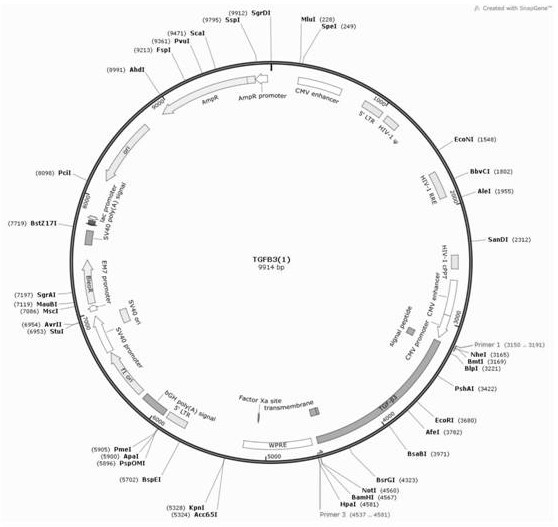
[0047] The construction of embodiment 1 lentiviral vector

[0048] 1. Design the gene sequence of the fusion protein TGF-β3. According to specific needs, the fusion protein we designed includes the N-terminal signal peptide, the target gene TGF-β3, the junction region and the transmembrane region. The structure of the N-terminal signal peptide and the transmembrane region adopts the corresponding structure of the surface marker CD44 of mesenchymal stem cells. The connecting region is a flexible chain of 8 amino acids, and the sequences of each part are as follows:

[0049] N-terminal signal peptide:

[0050] ATGGACAAGTTTTGGTGGCACGCAGCCTGGGGACTCTGCCTCGTGCCGCTGAGCCTGGCG (SEQ ID NO. 1)

[0051] Target gene TGF-β3:

[0052] ATGAAGATGCACTTGCAAAGGGCTCTGGTGGTCCTGGCCCTGCTGAACTTTGCCACGGTCAGCCTCTCTCTGTCCACTTGCACCACCTTGGACTTCGGCCACATCAAGAAGAAGAGGGTGGAAGCCATTAGGGGACAGATCTTGAGCAAGCTCAGGCTCACCAGCCCCCCTGAGCCAACGGTGATGACCCACGTCCCCTATCAGGTCCTGGCCCTTTACAACAGCACCCGGGAGCTGCTGGAGGAGATGCATGGGGAGA...

Embodiment 2

[0065] Example 2 Characterization of Mesenchymal Stem Cells Infected with TGF-β3 Virus

[0066] 1. Infect the cells with lentivirus, take 10ug of the target plasmid and add it to 950μL of 1×HBS, mix gently, and then slowly drop 50μL of CaCl into it 2 , mix gently and let stand for 20min, add to the HEK 293T cells with a density of 70%, mix gently, put in 37℃, 5%CO 2 After 12 hours, replace it with 10 mL of 30% FBS complete medium for culture. After 48 hours, take the cell supernatant, and centrifuge at 4000 rpm for 15 minutes at room temperature. Take the supernatant and add it to the mesenchymal stem cells with a cell density of 50%, add polybrene with a final concentration of 8ug / ml, mix well, and put it in 37°C, 5% CO 2 After 12 hours, replace with 10% FBS complete medium.

[0067] 2. Screen the mesenchymal stem cells that successfully express TGF-β3, add puromycin at a final concentration of 2ug / ml to the infected mesenchymal stem cells for screening, last for 2-3 days, ...

Embodiment 3

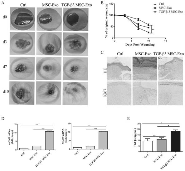
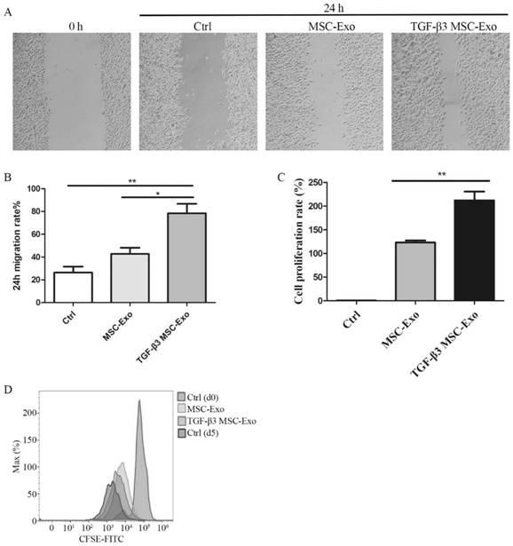
[0081] Example 3 Characterization of TGF-β3 exosomes secreted by mesenchymal stem cells infected with TGF-β3 lentivirus

[0082] 1. Extract exosomes from normal mesenchymal stem cells, mesenchymal stem cells co-incubated with TGF-β3 growth factor, and mesenchymal stem cells infected with TGF-β3 lentivirus. After the cells were cultured until the degree of confluence reached 50%, they were replaced with DMEM containing 0.5% fetal bovine serum and 1% P / S without exosomes and cultured for 48 hours. After the culture was completed, the cell culture medium was collected for gradient centrifugation and kept at 4°C throughout the process. 500×g, 10min, take the supernatant. 2000×g, 20min, take the supernatant. 10000×g, 40min, take the supernatant. The supernatant was centrifuged at 100,000×g for 90 min with an ultracentrifuge, and the precipitate was collected. After resuspending the pellet with PBS, continue to perform ultracentrifugation at 4°C, 100,000×g, 90 min, and take the ...


PUM

PropertyMeasurementUnit
Particle sizeaaaaaaaaaa

Abstract

The invention relates to a TGF-beta3 mesenchymal stem cell exosome and a preparation method and application thereof. The TGF-beta3 mesenchymal stem cell exosome is secreted by mesenchymal stem cells, and the TGF-beta3 mesenchymal stem cell exosome can express a TGF-beta3 fusion protein on a mesenchymal stem cell membrane; and the TGF-beta3 fusion protein, starting from the N-terminal, sequentially comprises an N-terminal signal peptide, a target TGF-beta3 protein, a connecting peptide and a mesenchymal stem cell transmembrane region. According to the invention, the obtained mesenchymal stem cell exosome capable of highly expressing TGF-beta3 on the membrane surface can play a role in promoting proliferation and migration of epidermal cells; and the TGF-beta3 mesenchymal stem cell exosome can not only retain the regeneration promoting capacity of mesenchymal stem cells, enhance the stability of TGF-beta3 and improve the specific targeting of the exosome to damaged skin cells, but also inhibit the activation and proliferation of immune cells, shorten the wound healing time and reduce scar formation, with a good treatment effect on tissue damage.

Description

technical field [0001] The present invention relates to the technical field of biomedicine, in particular to TGF-β3 mesenchymal stem cell exosomes and a preparation method and application thereof. Background technique [0002] Scarring and fibrosis are the end result of surgical and non-surgical skin injuries. Scarring and fibrosis not only lead to poor skin appearance but also impair skin function, sometimes causing adverse psychological effects and pain. Scar formation is associated with many It is related to various factors, such as the depth and size of the wound, the location of the injury, the difference in individual constitution, age and so on. Examples include severe scarring after burns, trauma and certain surgical procedures. Studies have found that animal embryo wounds show high levels of TGF-β3 and low levels of TGF-β1 and TGF-β2 expressions (Dev Biol.1991Sep; 147(1):207-15.doi:10.1016 / s0012-1606( 05)80018-1). It has also been proved in clinical experiments t...

Claims


Application Information

Patent Timeline
IPC IPC(8): C12N5/10C12N5/0775C12N15/62C12N15/867A61K35/28A61P17/02A61P29/00A61P43/00
CPCA61K35/28A61P17/02A61P29/00A61P43/00C07K14/495C07K2319/02C07K2319/03C12N5/0662C12N15/86C12N2740/15043
Inventor 陈玉容周晗邵静邹衡芳廖勇
Owner 广州远想生物科技股份有限公司
Why Eureka
Try Eureka