A kind of hybridoma cell secreting duck new reovirus σc protein monoclonal antibody, monoclonal antibody and application
A technology of reovirus and hybridoma cells, applied in the biological field, can solve the problem of unclear antigenicity information of NDRVσC protein epitope
- Summary
- Abstract
- Description
- Claims
- Application Information
AI Technical Summary
Problems solved by technology
Method used
Image

Examples
Embodiment 1
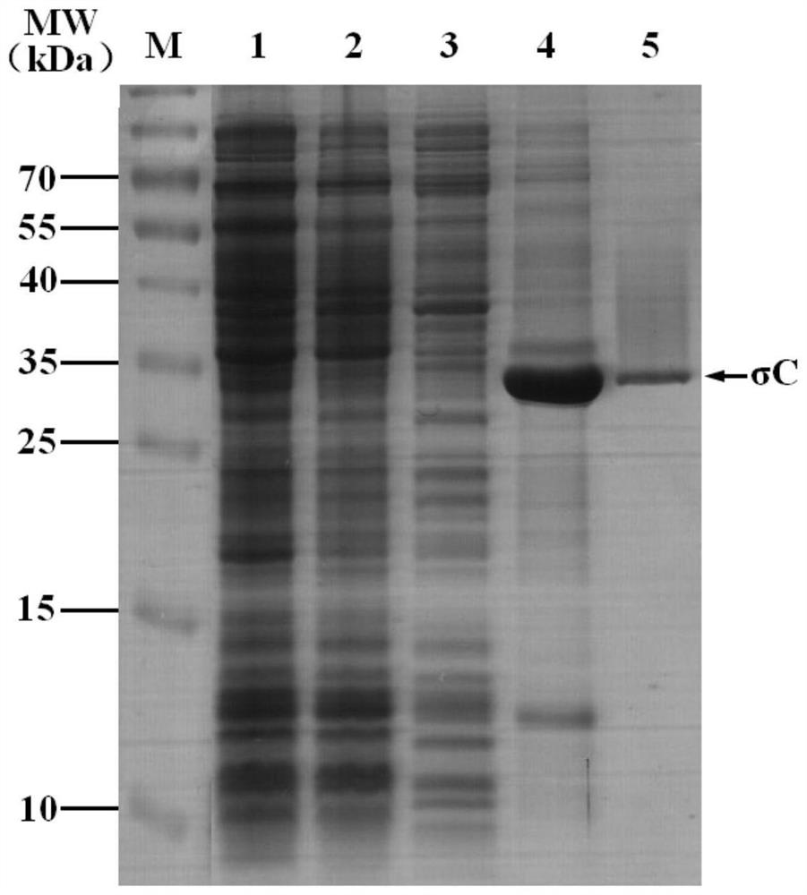
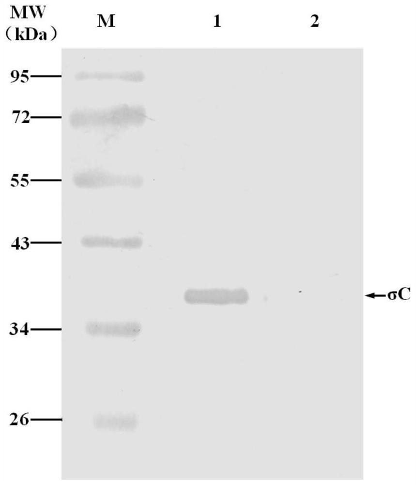
[0028] Example 1: Construction, protein expression and purification of recombinant plasmid pET-28a-σC
[0029] According to the S1 gene sequence (KF154116) of the NDRV ZJ00M strain, a pair of primers were designed with the σCORF of the S1 gene as the target region by using Oligo6.0 software, and the primers were synthesized by Invitrogen. Its sequence is as follows SigC-F: 5'-ACA CCATGG CGCAACGAGGTGATACGCCTG-3' (Nco I restriction site is underlined), SigC-R: 5'-ATA CTCG AG GCCCGTGGCGACGGTGAAGCGTAAC-3' (Xho I restriction site is underlined). The σC target fragment was amplified by RT-PCR, and the target fragment σC and the vector pET-28a were double-digested with Nco I / XhoI, respectively, and connected with T4 DNA ligase to construct a recombinant plasmid pET-28a-σC, which was transformed into the host bacteria In BL21, the expression was induced by IPTG, centrifuged, sonicated, and the supernatant and precipitate were separated. Expression of recombinant proteins was an...
Embodiment 2
[0031] Embodiment 2: Preparation of animal immunization, cell fusion and McAb
[0032] The purified recombinant σC protein was immunized by multi-point subcutaneous injection on the back of the neck and back of BALB / c mice aged 6-8 weeks (100 μg / mouse). Booster immunizations are given every two weeks. The first dose was emulsified with an equal volume of Freund's complete adjuvant, and the second and third doses were emulsified with an equal volume of Freund's incomplete adjuvant. Seven days after the third immunization, blood was collected from the tail, and the serum was separated and diluted in multiples, and the antibody titer of mouse serum was detected by ELISA plate coated with immunogen σC protein. The mice with the highest serum titer were boosted with σC protein (without adjuvant) (100 μg / mouse) 3 days before cell fusion. On the 3rd day, mouse spleen cells were taken for cell fusion with SP2 / 0 cells.
[0033] Fusion is carried out according to conventional methods...
Embodiment 3
[0036] Example 3: Identification of McAb biological properties
[0037] (1) McAb subclass identification
[0038] The McAb subtype identification kit was used to identify the subtype of the obtained MAbs. For specific steps, please refer to the instruction manual.
[0039] The results of antibody subtype identification showed that the antibody subtype secreted by the A5-B6 cell line was IgG1, and the secreted antibody light chains were all κ (Table 1).
[0040] Table 1 Subtype identification of hybridoma cell lines
[0041]
[0042] κ, λ: Antibody light chain class.
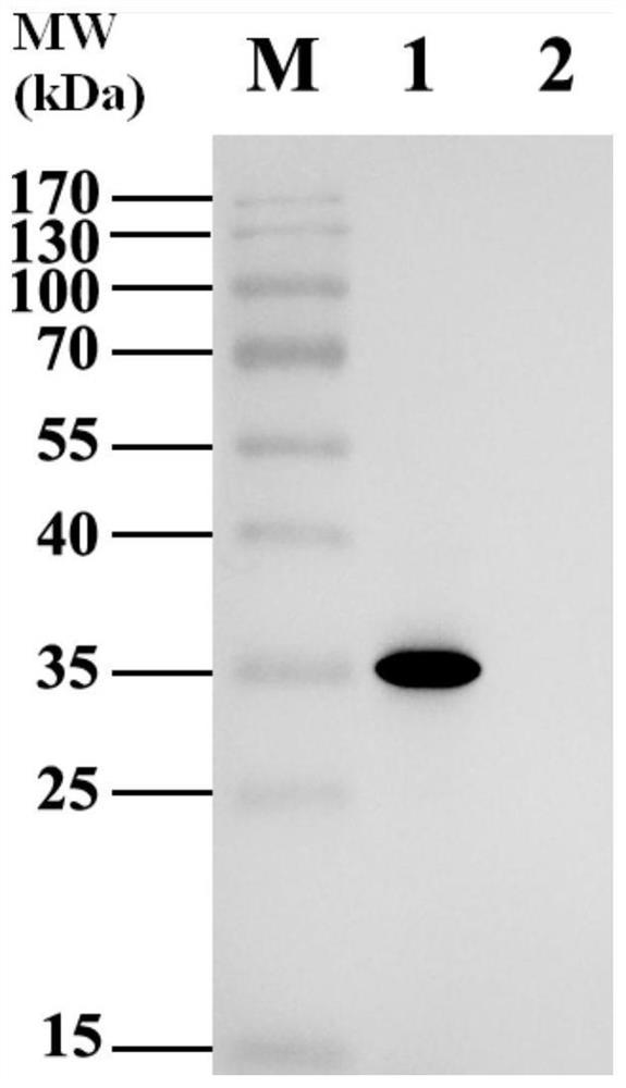
[0043] (2) Western blot analysis of McAb
[0044] Centrifuge the DF-1 cells infected with NDRV, discard the supernatant, add the sample buffer to the precipitate, and conduct SDS-PAGE electrophoresis after boiling. closed. The prepared A5-B6 monoclonal antibody was used as the primary antibody, goat anti-mouse IgG-HRP was used as the secondary antibody, and DF-1 cells not infected with NDRV were used as t...
PUM
 Abstract
Description
Claims
Application Information
 - R&D Engineer
- R&D Manager
- IP Professional
- Industry Leading Data Capabilities
- Powerful AI technology
- Patent DNA Extraction
Browse by: Latest US Patents, China's latest patents, Technical Efficacy Thesaurus, Application Domain, Technology Topic.
© 2024 PatSnap. All rights reserved.Legal|Privacy policy|Modern Slavery Act Transparency Statement|Sitemap